EV was extracted from 1 ml of plasma using a Size-Exclusion Chromatography column (qEV Original, 35nm, Izon Science) according to manufacturer's instructions.
Secondary antibody:
Cell Signaling 7074S (Rabbit)
No notes
Lysis Conditions:
SDS
at
95°C
for
7 min
Gel Conditions
Medium, 4 % acrylamide
Gel type:
BioRad 5671094
Run conditions:
120 V / 0 Amps for 60 min
Protein loading:
20 𝜇g
Reducing conditions:
β-mercaptoethanol
No notes
Transfer Conditions
Membrane type:
PVDF
Transfer method:
Semi-dry
Transfer buffer:
Tris-glycine with 20% methanol
Transfer voltage:
25 V
Transfer current:
2.5 Amps
Transfer temperature:
22°C
Transfer duration:
0.16 hr
Blocking
Blocking buffer:
0% milk, 0% BSA, No serum
Blocking temperature:
22°C
Duration:
1 hr
Primary Incubation
Antibody 1 dilution
1:1000
Incubation temperature
4°C
Incubation duration
16 hr
Number of washes
4
Wash buffer
TBST
Wash duration
10 min
Secondary Incubation
Antibody 2 dilution
1:1000
Incubation temperature
22°C
Incubation duration
1 hr
Number of washes
4
Wash buffer
TBST
Wash duration
10 min
Detection
Detection method
Enhanced chemiluminescence (ECL)
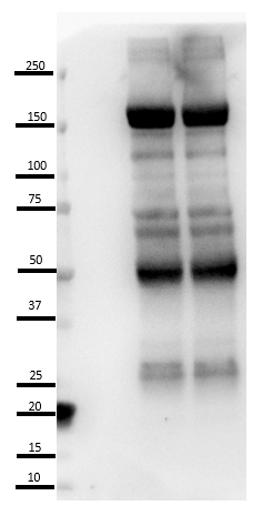
Expected band size
96 kDa
Result:
No specific bands
Background:
Multiple non-specific bands
Related Publications
–––
Weak or no signal is seen at the expected MW 96-100kda, whereas a non-specific strong signal is seen at 150kda
Weak or no signal is seen at the expected MW 96-100kda, whereas a non-specific strong signal is seen at 150kda