Registration and more information: 

ASIC Annual Meeting

When: October 17 – 19, 2024
Where: Bethesda North Marriott Hotel & Conference Center

Abstracts are due August 30, 2024
Registration deadline: September 25, 2024

The ASIC Annual Meeting is an intellectual “home” and support network for new and seasoned investigators, students, and postdocs. Over three days you’ll exchange ideas on emerging questions and cutting-edge developments in non-EV research including:

Extracellular Vesicles (EVs)
Extracellular Particles (EPs)
Particulate carriers of extracellular RNA (exRNA) as biological mediators, regulators, and diagnostic analytes.

The meeting also covers a broad range of intercellular communication processes including:

EVs
EPs
exRNA in Cancer
CNS Diseases
Infections (both bacterial and viral)

WHY ATTEND?
You’ll have a valuable opportunity to network with investigators from diverse scientific and clinical fields, discuss and advance the impact of EV/EP/ExRNA in diagnostics and treatments, and better understand the biogenesis of normal vs. disease states.

Are you new to the field? You can work with colleagues and mentors so you can improve your grants and develop their skills to become successful independent researchers.

Women and those from underrepresented groups are especially encouraged to attend and connect with seasoned researchers who have years of mentoring experience.

Thanks to Baylor College of Medicine for allowing us to share this press release here.
exRNA illustration, courtesy of Milosavljevic labexRNA / exRBP illustration
Figure Credit: Milosavljevic lab

An international team led by researchers at Baylor College of Medicine with the National Institutes of Health Extracellular RNA Communication Consortium and the Bogdan Mateescu laboratory at the ETH Zürich and University of Zürich has developed a powerful new resource to study extracellular RNA (exRNA), a novel form of cell-to-cell communication. The study, published in the journal Cell Genomics, lays the foundation to examine how exRNA and its carrier proteins found in bodily fluids function in a healthy as well as a diseased setting, potentially providing a means to accurately implement early detection and monitor disease processes.

Continue reading

This blog is a repost of the NIH Directors’ blog from April 4th, 2023.
Extracellular RNA Communication Consortium
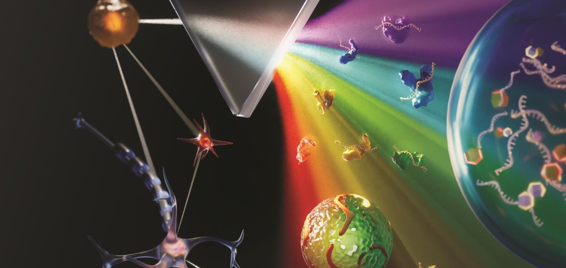
Figure Credit: XVIVO Scientific Animation, Wethersfield, CT

With just a blood sample from a patient, a promising technology has the potential to accurately diagnose non-small cell lung cancer (NSCLC), the most-common form of the disease, more than 90 percent of the time. The same technology can even predict from the same blood sample whether a patient will respond well to a targeted immunotherapy treatment.

This work is a good example of research supported by the NIH Common Fund. Many Common Fund programs support development of new tools that catalyze research across the full spectrum of biomedical science without focusing on a single disease or organ system.

The emerging NSCLC prediction technology was developed as part of our Extracellular RNA Communication Program. The program develops technologies to understand RNA circulating in the body, known as extracellular RNA (exRNA). These molecules can be easily accessed in bodily fluids such as blood, urine, and saliva, and they have enormous potential as biomarkers to better understand cancer and other diseases.

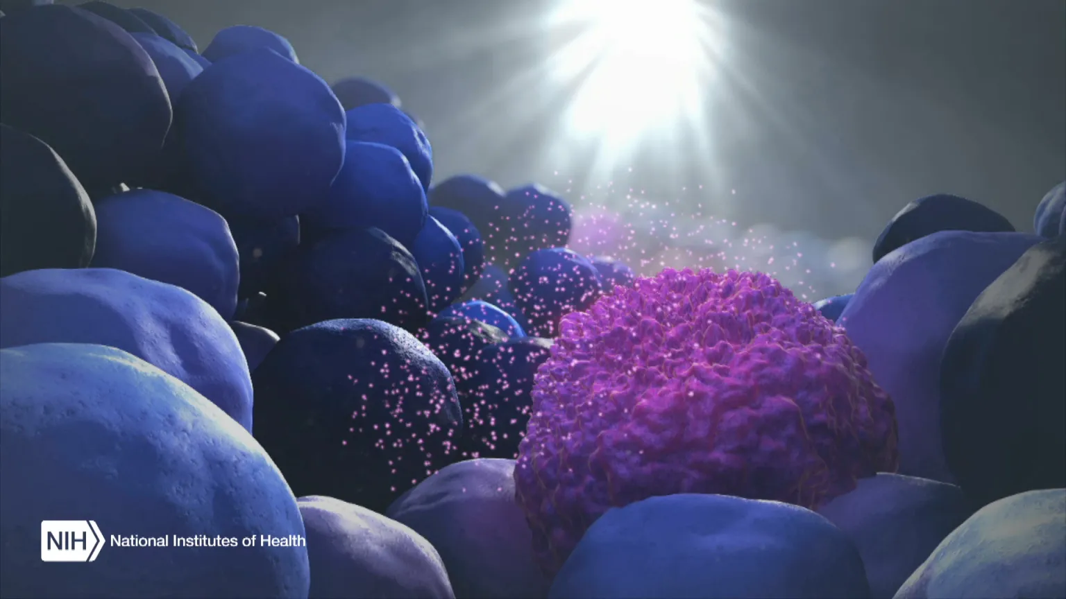
When the body’s immune system detects a developing tumor, it activates various immune cells that work together to kill the suspicious cells. But many tumors have found a way to evade the immune system by producing a protein called PD-L1.

Displayed on the surface of a cancer cell, PD-L1 can bind to a protein found on immune cells with the similar designation of PD-1. The binding of the two proteins keeps immune cells from killing tumor cells. One type of immunotherapy interferes with this binding process and can restore the natural ability of the immune system to kill the tumor cells.

However, tumors differ from person to person, and this form of cancer immunotherapy doesn’t work for everyone. People with higher levels of PD-L1 in their tumors generally have better response rates to immunotherapy, and that’s why oncologists test for the protein before attempting the treatment.

Because cancer cells within a tumor can vary greatly, a single biopsy taken at a single site in the tumor may miss cells with PD-L1. In fact, current prediction technologies using tissue biopsies correctly predict just 20 – 40 percent of NSCLC patients who will respond well to immunotherapy. This means some people receive immunotherapy who shouldn’t, while others don’t get it who might benefit.

To improve these predictions, a research team led by Eduardo Reátegui, The Ohio State University, Columbus, engineered a new technology to measure exRNA and proteins found within and on the surface of extracellular vesicles (EVs) [1]. EVs are tiny molecular containers released by cells. They carry RNA and proteins (including PD-L1) throughout the body and are known to play a role in communication between cells.

As the illustration above shows, EVs can be shed from tumors and then circulate in the bloodstream. That means their characteristics and internal cargo, including exRNA, can provide insight into the features of a tumor. But collecting EVs, breaking them open, and pooling their contents for assessment means that molecules occurring in small quantities (like PD-L1) can get lost in the mix. It also exposes delicate exRNA molecules to potential breakdown outside the protective EV.

The new technology solves these problems. It sorts and isolates individual EVs and measures both PD-1 and PD-L1 proteins, as well as exRNA that contains their genetic codes. This provides a more comprehensive picture of PD-L1 production within the tumor compared to a single biopsy sample. But also, measuring surface proteins and the contents of individual EVs makes this technique exquisitely sensitive.

By measuring proteins and the exRNA cargo from individual EVs, Reátegui and team found that the technology correctly predicted whether a patient had NSCLC 93.2 percent of the time. It also predicted immunotherapy response with an accuracy of 72.2 percent, far exceeding the current gold standard method.

The researchers are working on scaling up the technology, which would increase precision and allow for more simultaneous measurements. They are also working with the James Comprehensive Cancer Center at The Ohio State University to expand their testing. That includes validating the technology using banked clinical samples of blood and other bodily fluids from large groups of cancer patients. With continued development, this new technology could improve NSCLC treatment while, critically, lowering its cost.

The real power of the technology, though, lies in its flexibility. Its components can be swapped out to recognize any number of marker molecules for other diseases and conditions. That includes other cancers, neurodegenerative diseases, traumatic brain injury, viral diseases, and cardiovascular diseases. This broad applicability is an example of how Common Fund investments catalyze advances across the research spectrum that will help many people now and in the future.

Reference

[1] Nyugen LTH et al. An immunogold single extracellular vesicular RNA and protein (AuSERP) biochip to predict responses to immunotherapy in non-small cell lung cancer patients J Extracell Vesicles (2023) 11: e12258. doi: 10.1002/jev2.12258 PMID: 36093740.

Links

Extracellular RNA Communication Program (ERCC) (Common Fund)

Video: Unlocking the Mysteries of Extracellular RNA Communication

Upcoming Meeting: ERCC19 Research Meeting (May 1-2, 2023)

Eduardo Reátegui Group for Bioengineering Research (The Ohio State University College of Engineering, Columbus)

Note

Dr. Lawrence Tabak, who performs the duties of the NIH Director, has asked the heads of NIH’s Institutes, Centers, and Offices to contribute occasional guest posts to the blog to highlight some of the interesting science that they support and conduct. This is the 27th in the series of NIH guest posts that will run until a new permanent NIH director is in place.

This blog originated as a press release from the International Communications Office at Nagoya University. Thanks to them for allowing us to repost it here.

Researchers at Nagoya University in Japan have used a new device to identify a key membrane protein in urine that indicates whether the patient has a brain tumor. Their protein could be used to detect brain cancer, avoiding the need for invasive tests, and increasing the likelihood of tumors being detected early enough for surgery. This research could also have potential implications for detecting other types of cancer. The research was published in ACS Nano.

Continue reading

This blog originated as a press release from Hokkaido University. Thanks to them for allowing us to repost it here.

Researchers from Hokkaido University and Toppan have developed a method to detect build-up of amyloid β in the brain, a characteristic of Alzheimer’s disease, from biomarkers in blood samples.

Alzheimer’s disease is a neurodegenerative disease, characterised by a gradual loss of neurons and synapses in the brain. One of the primary causes of Alzheimer’s disease is the accumulation of amyloid β (Aβ) in the brain, where it forms plaques. Alzheimer’s disease is mostly seen in individuals over 65 years of age, and cannot currently be stopped or reversed. Thus, Alzheimer’s disease is a major concern for nations with ageing populations, such as Japan.

A team of scientists from Hokkaido University and Toppan, led by Specially Appointed Associate Professor Kohei Yuyama at the Faculty of Advanced Life Science, Hokkaido University, have developed a biosensing technology that can detect Aβ-binding exosomes in the blood of mice, which increase as Aβ accumulates in the brain. Their research was published in the journal Alzheimer’s Research & Therapy.

Continue reading

Cells can communicate with one another to coordinate essential processes such as development, growth, and repair through the release of signaling intermediates. One class of signaling intermediates are extracellular vesicles (EVs) that contain nucleic acids and proteins that mediate cell-cell communication. In cancer, the cargo of these EVs is altered in order to promote tumor progression, improving the ability to proliferate, invade, metastasize, and develop drug resistance, among other cancer characteristics. While most EVs range in diameter from 50 nanometers to one micron, there has been an increasing interest in smaller particles that might also be released from cells and contribute to cancer. Only recently has technology evolved enough to detect these previously undiscernible nanoparticles. Qin Zhang, PhD, Robert Coffey, MD, and colleagues were motivated by previous advances in the lab regarding the role of EVs in cancer to determine if smaller particles existed with similar functions. Dr. Coffey and his team discovered a new nanoparticle, termed the supermere, with functional relevance not only to cancer but to many other diseases, resulting in a publication at the end of 2021 in Nature Cell Biology (Zhang Q et al. 2021).

Continue reading

This blog originated as a press release from Nagoya University. Thanks to them for allowing us to repost it here.

Researchers at Nagoya University in Japan have developed a new chemical-only process that may represent an important breakthrough in creating customized mRNA vaccines for a variety of diseases and allow for the inexpensive preparation of mRNA in large quantities.

During the COVID-19 pandemic, mRNA vaccines were successfully used to boost immunity. These vaccines teach cells how to make a protein that triggers the body’s immune response, allowing its natural defenses to recognize the invading virus. However, current vaccines that use biological processes do not allow for the precise molecular design of mRNA, which limits their use in creating new vaccines as variants emerge.

As published in ACS Chemical Biology, a research group led by Professor Hiroshi Abe and Associate Professor Naoko Abe of the Graduate School of Science at Nagoya University has developed the first completely chemical synthesis method for mRNA.

In their study, the group synthesized a part of the mRNA called the cap. The cap is important because it promotes the translation of mRNA into proteins and protects mRNA from degradation. To prepare synthetic mRNA, such as that used in vaccines, the two currently used biological methods rely on enzymes to incorporate the cap structure into the mRNA. However, the researchers found that their technique could synthesize a variety of chemically modified mRNA strands with a cap structure.

According to Professor Abe, “our research suggests that it is possible to make mRNAs with precisely introduced chemical modifications with complete control over the process. The molecular design reported in our study exhibits five times higher translational activity than that of enzyme-produced natural-type mRNA. This means that mRNA can be synthesized in large quantities at low cost using chemical synthesis.”

Chemically modified mRNA could be used to create customized vaccines against a variety of infectious diseases including viruses and cancers. Professor Abe explains, “By introducing these chemical modifications, the mRNA becomes stable. This could allow for the creation of long-lasting and effective mRNA vaccines. In addition, it could allow mRNA to be administered directly instead of using lipid nanoparticles, which are used for delivery in current vaccines.”

“One of the exciting implications of this research is that this could be used in the next generation of vaccines,” the researchers said. “We hope that the capping method reported here will be of great use in the development of RNA therapeutics.”

The study, “Complete Chemical Synthesis of Minimal Messenger RNA by Efficient Chemical Capping Reaction,” was published in ACS Chemical Biology on May 24, 2022.

Authors:
Naoko Abe, Akihiro Imaeda, Masahito Inagaki, Zhenmin Li, Daisuke Kawaguchi, Kaoru Onda, Yuko Nakashima, Satoshi Uchida, Fumitaka Hashiya, Yasuaki Kimura, and Hiroshi Abe

The study was supported by the AMED LEAP project ‘Innovation of Chemistry-Based Molecular Design and Production Methods for mRNA and its Application to Vaccines’, which started in FY2021.

Reference

Abe N et al. Complete chemical synthesis of minimal messenger RNA by efficient chemical capping reaction. ACS Chem Biol AOP 2022-05-24. doi: 10.1021/acschembio.1c00996 PMID: 35608277.

This blog originated as a press release from the Johns Hopkins Kimmel Cancer Center. Thanks to them for allowing us to repost it here.

It may be possible to identify the presence of an aggressive brain tumor in children by studying their cerebrospinal fluid, according to new research led by Johns Hopkins Kimmel Cancer Center investigators.

Comparing cerebrospinal fluid samples from 40 patients with medulloblastoma — the most common malignant brain tumor in children, accounting for 10% to 15% of pediatric central nervous system tumors — and from 11 healthy children without the disease, investigators identified 110 genes, 10 types of RNA–the machinery that translates proteins–called circular RNAs, 14 lipids and several metabolites that were expressed differently between the two groups. While these details were not specific enough to distinguish among the four subtypes of medulloblastoma, they could be used to identify the presence of cancer versus normal fluid. A description of the work was published in the journal Acta Neuropathologica Communications.

Continue reading

Extracellular vesicles (EVs) are small membrane-bound particles that are loaded with various proteins, RNA, DNA, and lipids, and secreted by cells. Interest in these vesicles has grown in recent years with mounting evidence that EVs act as intercellular communication systems, transferring their selected cargo to other cells to confer specific effects on target cell biology. However, the processes that direct specific RNAs and proteins into these specialized vesicles remain largely unknown. In the April 25th, 2022 edition of Developmental Cell, Alissa Weaver, M.D., Ph.D. and her research team at the Vanderbilt Center for Extracellular Vesicle Research have uncovered subcellular hubs of EV formation that selectively assemble RNA-containing EVs. These hubs are located at membrane contact sites (MCS) where the endoplasmic reticulum (ER) interacts with EV biogenesis membranes, including late endosomal multivesicular bodies (MVBs) (ER-MVB MCS). Shedding much needed mechanistic light, the group further pinpointed the ER MCS membrane tether protein VAP-A and its binding partner ceramide transfer protein (CERT) as key drivers in this process.

Continue reading

This blog originated as a press release from the Swiss National Centre of Competence in Research SYNAPSY. Thanks to them for allowing us to repost it here.

A research team from Synapsy has shown that the severity of the clinical symptoms of schizophrenia is strongly linked to blood biomarkers related to the deregulation of neuronal mitochondria.

Psychotic symptoms are a characteristic clinical manifestation of schizophrenia. They go hand-in-hand with an increase in oxidative stress, which results in damage to a particular type of neurons called parvalbumin neurons. This deterioration leads to dysfunction in the activity of the prefrontal cortex, a region of the brain that is involved in cognition. A study conducted at the Centre for Psychiatric Neuroscience of the Lausanne University (UNIL) and the Lausanne University Hospital (CHUV), and supported by the National Centre of Competences in Research Synapsy (Synapsy), has shown, in an animal model, that the cellular mechanism for recycling mitochondria is deficient in parvalbumin neurons. The study – published in the journal Molecular Psychiatry – investigated the underlying biochemical mechanisms, pinpointing two key molecules, miR-137 and COX6A2, that can be detected in blood. When used as biomarkers in patients diagnosed with psychosis, they unveil two distinct clinical sub-groups with different severity of symptoms, cognitive deficits, and functioning in everyday life. This discovery represents a major breakthrough for stratifying individuals suffering from schizophrenia, whose heterogeneity of symptoms currently restricts diagnosis and treatment.

Mouse parvalbumin neuronsMouse parvalbumin neurons – Parvalbumin neurons are affected by oxidative stress in mouse model of schizophrenia. Markers of this neuropathological process can be detected in the blood of human patients, helping to diagnose and potentially treat them with antioxidant compounds. © Inès Khadimallah / CHUV UNIL.

Continue reading