Year: 2017

(This blog first appeared as a press release from Ohio State University.)

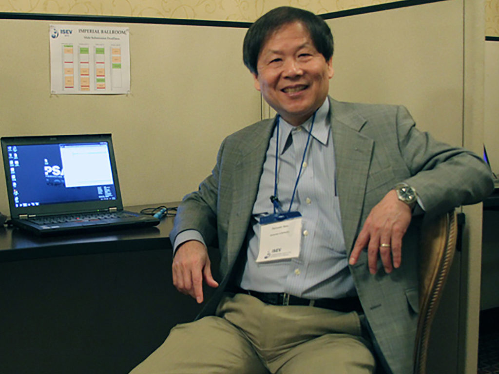
Principal investigator Peixuan Guo, PhD, Sylvan G. Frank Endowed Chair professor of the OSU College of Pharmacy and a member of the OSUCCC – James Translational Therapeutics Program.

  • Therapies based on RNA, such as small interfering RNA, hold great promise for cancer treatment but delivering these agents to their targets in cancer cells has been a problem.
  • A new study shows that attaching antibody-like RNA nanoparticles to microvesicles can deliver effective RNA therapeutics specifically to cancer cells.
  • The researchers are now working to adapt the technology for use in the clinic.

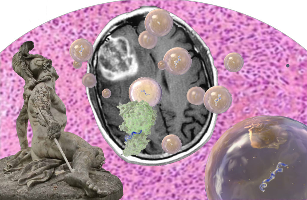
Columbus, Ohio – A new study shows that attaching antibody-like RNA nanoparticles to microvesicles can deliver effective RNA therapeutics such as small interfering RNA (siRNA) specifically to cancer cells. Researchers used RNA nanotechnology to apply the RNA nanoparticles and control their orientation to produce microscopic, therapy-loaded extracellular vesicles that successfully targeted three types of cancer in animal models.

The findings, reported in the journal Nature Nanotechnology, could lead to a new generation of anticancer drugs that use siRNA, microRNA and other RNA-interference technologies.

The study was led by researchers at Ohio State’s College of Pharmacy; the Ohio State University Comprehensive Cancer Center – James Cancer Hospital and Solove Research Institute (OSUCCC – James).

“Therapies that use siRNA and RNA interference technologies are poised to transform cancer therapy,” says the principal investigator Peixuan Guo, PhD, Sylvan G. Frank Endowed Chair professor of the College of Pharmacy and a member of the OSUCCC – James Translational Therapeutics Program. “But clinical trials evaluating these agents have failed one after another due to the inability to deliver the agents directly to cancer cells in the human body.”

Guo noted that even when agents did reach and enter cancer cells, they were trapped in internal vesicles called endosomes and rendered ineffective.

“Our findings solve two major problems that impede these promising anticancer treatments: targeted delivery of the vesicles to tumor cells and freeing the therapeutic from the endosome traps after it is taken up by cancer cells. In this study, cancers stopped growing after systemic injection of these particles into animal models with tumors derived from human patients.” Guo says. “We’re working now to translate this technology into clinical applications.”

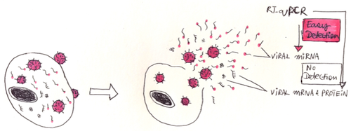
Guo and his colleagues produced extracellular microvesicles (exosomes) that display antibody-like RNA molecules called aptamers that bind with a surface marker that is overexpressed by each of three tumor types:

  • To inhibit prostate cancer, vesicles were designed to bind to prostate-specific membrane antigen (PSMA);
  • To inhibit breast cancer, vesicles were designed to bind to epidermal growth factor receptor (EGFR);
  • To inhibit a colorectal cancer graft of human origin, vesicles were designed to bind to folate receptors.

All vesicles were loaded with a small interfering RNA for down-regulating the survivin gene as a test therapy. The survivin gene inhibits apoptosis and is overexpressed in many cancer types.

Key findings include:

  • Vesicles targeting the prostate-specific membrane antigen completely inhibited prostate-cancer growth in an animal model with no observed toxicity.
  • Vesicles targeting EGFR inhibited breast cancer growth in an animal model.
  • Vesicles targeting folate receptors significantly suppressed tumor growth of human patient-derived colorectal cancer in an animal model.

“Overall, our study suggests that RNA nanotechnology can be used to program natural extracellular vesicles for delivery of interfering RNAs specifically to cancer cells,” Guo says.

Funding from the National Institutes of Health/National Cancer Institute (grants TR000875 and CA207946, CA186100, CA197706, CA177558 and CA195573) supported this research.

Other researchers involved in this study were Fengmei Pi, Daniel W. Binzel, Zhefeng Li, Hui Li, Farzin Haque, Shaoying Wang and Carlo M. Croce, The Ohio State University Wexner Medical Center; Meiyan Sun and Bin Guo, University of Houston; Piotr Rychahou and B. Mark Evers, University of Kentucky; and Tae Jin Lee, now at University of Texas.

About the OSUCCC – James
The Ohio State University Comprehensive Cancer Center – Arthur G. James Cancer Hospital and Richard J. Solove Research Institute strives to create a cancer-free world by integrating scientific research with excellence in education and patient-centered care, a strategy that leads to better methods of prevention, detection and treatment. Ohio State is one of only 49 National Cancer Institute (NCI)-designated Comprehensive Cancer Centers and one of only a few centers funded by the NCI to conduct both phase I and phase II clinical trials on novel anticancer drugs sponsored by the NCI. As the cancer program’s 308-bed adult patient-care component, The James is one of the top cancer hospitals in the nation as ranked by U.S. News & World Report and has achieved Magnet designation, the highest honor an organization can receive for quality patient care and professional nursing practice. At 21 floors with more than 1.1 million square feet, The James is a transformational facility that fosters collaboration and integration of cancer research and clinical cancer care.

Quantitative measurements of the number, size, and cargo of extracellular vesicles (EVs) are essential to both basic research on how EVs are produced and function, and to application of this knowledge to the development of EV-based biomarkers and therapeutics. Flow cytometry is a popular method for analyzing EVs, but their small size and dim signals have made this a challenge using the conventional flow cytometry approaches developed for analysis of cells (1). Moreover, established flow cytometry calibrators, standards, and experimental design considerations for cell studies are not regularly used in EV studies. As a result, there is significant variation in instrument set up, sample preparation, and data reporting for flow cytometric measurements of EVs. These issues are increasingly appreciated (1-5), but much needs to be done to develop consensus on best practices. To address these issues, members of the International Society for Extracellular Vesicles (ISEV), the International Society for Advancement of Cytometry (ISAC), and the International Society on Thrombosis and Hemostasis (ISTH) are participating in a tri-Society Working Group, which includes several ERCC members, to improve the reporting of methods and results for FC-based EV measurements.

Reporting of EV Measurement Methods

A flow cytometer is an instrument, not a method. An EV analysis method that uses a flow cytometer involves many instrument setup, sample preparation, and data analysis decisions, including: 1) what signal to use for EV detection (light scatter or fluorescence); 2) how to resolve single EVs from the simultaneous occurrence of many EVs in the laser at the same time (aka coincidence or “swarm”); 3) how to gate the data to focus on EVs versus background events (without introducing artifacts or mis-representing the data); 4) how to estimate the size of the particles detected; 5) how to estimate the brightness of the particles detected; 6) how to verify that the particles detected are EVs and not other particles present in the sample, to name just some of the many decisions involved.

Several years ago, ISAC developed and introduced the Minimum Information about a Flow Cytometry Experiment (MIFlowCyt) (6), a set of guidelines to promote the sharing, reproducibility, and proper interpretation of flow cytometry data. These guidelines were developed with cell analysis, and particularly high parameter immunophenotyping, in mind, but they also apply to multiparameter EV analysis. However, there are several additional details about an EV measurement that are essential to include. The ISEV-ISAC-ISTH EV FC Working Group has been conducting a series of standardization studies to develop a consensus on the essential elements of an FC-based EV measurement that should be reported. These studies will be reported, along with the consensus reporting guidelines, in a paper planned for the coming year.

Standards and Calibrators for EV Analysis

Standards and calibration are essential components of any analytical method. These standards, and their use, are well established for flow cytometry and include 1) counting beads that can be used to calibrate sample flow rates for reporting of absolute particle concentrations, 2) fluorescence intensity standards that enable particle brightness to be expressed in NIST-traceable absolute units of mean equivalent soluble fluorochromes (MESF) (7) or equivalent reference fluorochromes (ERF) (8); 3) antibody-capture standards that can be used to estimate antibody binding in immunofluorescence measurements; and 4) NIST-traceable particle size standards.

Most of these standards and calibrators, and their methods of use, can be applied to EV measurements, with some caveats and cautions. Particle size standards, in particular, are often mis-used in FC-based EV measurements due to a lack of understanding of the effect on light scatter of refractive index (RI), which is different for polystyrene, silica, and lipids. With care, however, these differences can be used in conjunction with Mie scattering theory to enable estimates of EV size based on FC light scatter measurements. Commercially available fluorescence intensity and antibody-capture standards are generally designed for cell measurements, and tend to be brighter than EVs, but still have value for facilitating comparison of measurements between labs or instruments. EV-scaled intensity and antibody-binding standards will be a useful addition to the EV analysis toolbox, and are in development by several groups and companies.

A major unmet need is for EV standards, which will have use not only in FC-based EV measurements, but across the EV field. This is a challenging prospect, as an ideal EV standard will reflect not only the size and number of EVs, but also cargo, including surface molecules (for immunophenotyping) and intra-vesicular cargo, including nucleic acids, soluble proteins, and small molecules. Moreover, EVs are themselves quite diverse, raising the question of what type of EV, if any, might represent a universal standard. EV preparations for various cultured cell lines are commercially available from a number of sources but, in general, these have not been subjected to rigorous, independent characterization of these essential features or their uniformity, stability, or reproducibility. Such characterization is essential for validation of any putative standard and may be the subject of future activities by the ISEV-ISAC-ISTH EV FC Working Group.

Conclusions and Prospects

As EV research expands to impact every area of biology, issues with rigor and reproducibility are front and center. Translating observations made in the basic research lab into mechanistic understanding of EV actions and clinically actionable knowledge requires robust and validated analytical methods. Careful attention to the description of methods, standardization and calibration of analytical instrument and methods, and reporting of results are essential. Community efforts by the ERCC and relevant international societies will be key to helping researchers maximize the value of their work to the broader community.

In future blog posts we will discuss the controversial issue of whether to use light scatter or fluorescence to detect EVs, as well as new EV detection methods we’ve developed using fluorogenic membrane probes.

References

1. Nolan JP. Flow cytometry of extracellular vesicles: potential, pitfalls, and prospects. Curr. Protoc. Cytom. (2015) 73:13.14.1-13.14.16. PMID: 26132176. doi: 10.1002/0471142956.cy1314s73.
2. Chandler WL. Measurement of microvesicle levels in human blood using flow cytometry. Cytometry B Clin. Cytom. (2016) 90:326-336. PMID: 26606416. doi: 10.1002/cyto.b.21343.
3. Coumans FA, et al. Methodological guidelines to study extracellular vesicles. Circ. Res. (2017) 120:1632-1648. PMID: 28495994. doi: 10.1161/CIRCRESAHA.117.309417.
4. Nolan JP, Duggan E. Analysis of individual extracellular vesicles by flow cytometry. Methods Mol. Biol. (2018) 1678:79-92. PMID: 29071676. doi: 10.1007/978-1-4939-7346-0_5.
5. Nolan JP, Jones JC. Detection of platelet vesicles by flow cytometry. Platelets (2017) 28:256-262. PMID: 28277059. doi: 10.1080/09537104.2017.1280602.
6. Lee JA, et al. MIFlowCyt: the minimum information about a flow cytometry experiment. Cytometry A. (2008) 73:926-930. PMID: 18752282 doi: 10.1002/cyto.a.20623.
7. Wang L, Gaigalas AK, Abbasi F, Marti GE, Vogt RF, Schwartz A. Quantitating fluorescence intensity from fluorophores: practical use of MESF values. J. Res. Natl. Inst. Stand. Technol. (2002) 107:339-354. PMID: 27446735. doi: 10.6028/jres.107.027.
8. Wang L, Gaigalas AK. Development of multicolor flow cytometry calibration standards: Assignment of equivalent reference fluorophores (ERF) unit. J. Res. Natl. Inst. Stand. Technol. (2011) 116:671-83. PMID: 26989591. doi: 10.6028/jres.116.012.


Scientists from the ERCC have joined forces to create a CSF Consortium to pool resources and establish standard practices in the study of cerebrospinal fluid (CSF).

One of the goals of the ERCC is not only to understand the fundamental biology of extracellular RNA (exRNA), but to develop exRNA-based biomarkers of disease. When such biomarkers have been found, studied, and cleared for clinical use, liquid biopsy of blood and other biofluids can enable earlier disease detection and less invasive tracking of disease progression. For neurological disorders, drawing CSF from the spinal cord has clear benefits over a more invasive brain biopsy. Progress in our technical understanding of how to accurately assess biomarkers in CSF will increase our basic understanding and promote clinical advancements in the diagnosis and treatment of neurological disease. Unfortunately, there are many inconsistencies between the processing of CSF in current studies. Data replication is often difficult, in large part due to variability across laboratories and institutions in protocols for sample isolation, purification, and analysis. Thus, the CSF Consortium, spearheaded by Dr. Fred Hochberg (https://fredhhochbergmd.com), was designed to be a resource for researchers to help minimize these discrepancies.

The CSF consortium plan calls for CSF researchers and clinicians to work together to improve standard practices. A major focus is transparency through open sharing of their work. Researchers are encouraged to establish collaborations, share in-depth details of experimental designs and reagents (including batch/lot numbers), and release any details of in-house protocol modifications. Working with the same biosamples shared through the Virtual Biorepository (VBR) enables multiple labs to compare and synchronize their protocols with one source of variability removed. The expectation is that sharing of detailed information will enable future researchers to avoid common pitfalls and plan their own experiments appropriately. Ultimately, the goal is to have open-access information available from each stage of every project: from biofluid, RNA, and extracellular vesicle (EV) collection, isolation, and storage to downstream analyses such as RT-qPCR and RNA sequencing.

If you are a CSF researcher, please contact us so that we can work with you as well!

Highlights of recent CSF Consortium efforts
Saugstad et al. (2017) recently demonstrated the strength of the CSF consortium. In a collaboration between three institutions (UC San Diego, Oregon Health & Science University, and the Translational Genomics Research Institute), researchers worked together to characterize the EV and RNA composition of identical pools of CSF at each institute from patients with five different neurological disorders. This work in parallel allowed the groups to identify potential sources of variability in protocols including sample preparation, RNA isolation, and quantification of RNA via RNA sequencing and RT-qPCR. The study identified changes in EVs and RNA in the disease CSF samples and detected an enrichment of microRNAs and mRNAs related to disease in both EV and total RNA. The paper highlights the importance of stringent standard operating procedures, including the use of common standard sample collection and data analysis protocols across institutions.

In other work, Figueroa et al., 2017 performed a multi-institutional study of RNA extracted from CSF-derived EVs of patients with glioblastoma (GBM), a very aggressive form of brain cancer. (See this related blog on glioblastoma.) A key diagnostic biomarker in classical GBM is the functional status of the Epidermal Growth Factor Receptor (EGFR). This cell-surface receptor is the starting point of a series of signaling pathways related to cell growth. When its expression surges or it folds incorrectly, the result is cells with hyper-active signaling that never stop growing. This study involved the development of a liquid biopsy that scans RNA extracted from CSF EVs for tumor-associated amplifications and mutations in EGFR. The test has very high specificity and fair sensitivity: it almost never incorrectly flags a healthy patient as having GBM and correctly identifies almost two thirds of GBM sufferers. The clinical standard for diagnosis of GBM is magnetic resonance imaging (MRI), which correctly classifies most brain tumors, but in too many cases incorrectly suggests that healthy brain tissue might be cancerous. The complementarity of highly sensitive MRI and highly specific RNA liquid biopsy argues that updating the standard of care to include collection of CSF and brain images at the same time would better separate healthy from diseased brain tissue.

The CSF Consortium is casting its nets wider in its fight against glioblastoma, looking at molecules beyond EGFR in the attempt to develop an RNA-based diagnosis tool for GBM. Akers et al., 2017 developed a diagnostic panel of 9 miRNA biomarkers by analyzing the EV RNA from 135 CSF samples in 3 cohorts, followed by validation of the miRNA panel in 60 CSF samples from 2 cohorts. The researchers found that even with that fairly large sample size, the miRNA profiles in lumbar and cisternal CSF — fluid collected from the spine or the base of the neck, respectively — are significantly different, which is problematic, since cisternal CSF is much more difficult to collect. On the other hand, they also found that RNAs extracted from raw CSF had a similar profile and diagnostic power as RNAs extracted from vesicles after an initial EV purification step, which might simplify the translation of this biomarker research into the clinic.

References
Akers, J.C. et al. A cerebrospinal fluid microRNA signature as biomarker for glioblastoma. Oncotarget (2017) 8: 68769-68779.
Figueroa, J.M et al. Detection of wtEGFR amplification and EGFRvIII mutation in CSF-derived extracellular vesicles of glioblastoma patients. Neuro. Oncol. (2017) Advance online publication. doi: 10.1093/neuonc/nox085
Saugstad, J.A. et al. Analysis of extracellular RNA in cerebrospinal fluid. J. Extracellular Vesicles (2017) 6: 1317577.

We are pleased to announce the publication of miRandola 2017 in Nucleic Acids Research, Database issue 2018!
Citation:
miRandola 2017: a curated knowledge base of non-invasive biomarkers.
Francesco Russo*, Sebastiano Di Bella, Federica Vannini, Gabriele Berti, Flavia Scoyni, Helen V. Cook, Alberto Santos, Giovanni Nigita, Vincenzo Bonnici, Alessandro Laganà, Filippo Geraci, Alfredo Pulvirenti, Rosalba Giugno, Federico De Masi, Kirstine Belling, Lars J. Jensen, Søren Brunak, Marco Pellegrini, Alfredo Ferro.

*Correspondence to francesco.russo@cpr.ku.dk
URL: Nucleic Acids Research
Website: https://mirandola.iit.cnr.it/

The Extracellular RNA Communication Consortium (ERCC) has developed a Virtual Biorepository (VBR) to facilitate the sharing of biological materials between researchers. As of September 1, 2017, the VBR hub is now available for use by the global extracellular RNA research community (both ERCC and non-ERCC members) at https://genboree.org/vbr-hub. This Phase 1 (beta) release currently provides access to metadata on more than 10,000 biosamples. Specifically, there are 7,651 cerebrospinal fluid (CSF) and 2,356 hepatobiliary samples from the Translational Genomics Research Institute, Phoenix Children’s Hospital, Oregon Health and Science University, and the University of California, San Diego. Another 50,000 hepatobiliary samples from the Mayo Clinic are planned to be available before the end of 2017. Most participant institutions have agreed to a common framework for biosample exchange, including common Institutional Review Board (IRB) protocols and Material Transfer Agreements (MTA).

The Virtual Biorepository originally arose from the needs of investigators within the ERC consortium to share biofluid samples across institutions for the purpose of collaborative protocol development and biomarker discovery. To enable efficient sample sharing, the ERCC Resource Sharing Working Group worked with the Data Coordination Center (DCC) and Administrative Core to initiate VBR development. The initial goal was to enable the sharing of cerebrospinal fluid (CSF) samples among members of the ERCC-based CSF consortium. The types of shared resources available in the VBR have since extended to include hepatobiliary samples, tissue, cell, and macromolecular samples, and even sample slides. These resources may be useful for catalyzing collaborations during the next stage of the Extracellular RNA Communication project.

The VBR is a distributed database system consisting of a hub and a set of local or cloud-hosted nodes. The VBR hub provides an overview of the types and number of biosamples present at the nodes. The hub supports sample queries based on consortium (CSF, hepatobiliary), institution, and on publicly shared metadata about anonymized VBR biosamples, including clinical, radiographic, pathologic, and accession metadata. Lists of samples that satisfy search criteria are placed in a shopping cart for ordering from sample providers. Search criteria and results can be saved for later retrieval and modification. In the current implementation phase (Phase 1) of the biorepository, after selecting samples, researchers communicate directly with each other to make specific arrangements for sharing biosamples. Future improvements (Phase 2) of the shopping cart feature will allow end-to-end tracking of the biosample ordering and exchange process.

VBR nodes are set up independently of the hub and are under the control of sample providers. The ERCC DCC provides assistance regarding maintenance of data within individual VBR nodes using pre-defined metadata templates. Investigators potentially interested in setting up a VBR node to share metadata about their samples may contact the VBR administrator (thistlew@bcm.edu).

Thanks to Laurence de Nijs and the European College of Neuropsychopharmacology (ENCP) for allowing us to adapt their press release into a blog.


Individuals affected with PTSD (Post-Traumatic Stress Disorder) demonstrate changes in microRNA (miRNA) molecules associated with gene regulation. A controlled study, involving Dutch military personnel on deployment to a combat zone in Afghanistan, provided evidence for the role of blood-based miRNAs as candidate biomarkers for symptoms of PTSD. This finding may offer an approach towards screening for symptoms of PTSD, and it holds promise for understanding other trauma-related psychiatric disorders. Limitations of the study are that this was a small pilot study, and the findings need to be validated, extended, and confirmed. First results were presented at the 30th conference of the European College of Neuropsychopharmacology (ENCP) in Paris in early September.

PTSD is a psychiatric disorder which can manifest following exposure to a traumatic event, such as combat, assault, or natural disaster. Among individuals exposed to traumatic events, only a minority of individuals will develop PTSD, while others will show resiliency. Little is known of the mechanisms behind these different responses. The last few years have seen much attention given to whether the modification and expression of genes – epigenetic modifications – might be involved. But there are several practical and ethical challenges in designing a research study on humans undergoing such experiences, meaning that designing relevant study approaches is difficult.

A research group from the Netherlands worked with just over 1,000 Dutch soldiers and the Dutch Ministry of Defense to study changes in biology in relation to changes in presentations of symptoms of PTSD in soldiers who were deployed to a combat zone in Afghanistan. In a longitudinal study, they collected blood samples before deployment as well as 6 months after deployment. Most of the soldiers had been exposed to trauma, and some of the soldiers had developed symptoms of PTSD.

For this pilot study, from the initial group, 24 subjects were selected in 3 subgroups of 8. Eight of the soldiers had developed symptoms of PTSD; 8 had endorsed traumatic experiences but had not developed symptoms of PTSD; and another 8 had not been in serious traumatic circumstances and served as a control group. Using modern sequencing techniques, several types of miRNAs for which the blood levels differed between the groups were identified.

MiRNAs (Micro RiboNucleic Acids) are small molecules with chemical building blocks similar to DNA. Unlike the more famous DNA, miRNAs are typically very short – comprising only around 20 to 25 base units (the building blocks of nucleic acids), and they do not code, in other words they do not specify the production of a protein or peptide. However, they have very important roles in biology (every miRNA regulates the expression, and thereby also the activity of several other genes), and they are known to regulate the impact of environmental factors on biology. In addition, brain-derived miRNA can circulate throughout the human body and can be detected in the blood.

Differences in miRNA levels have been associated with certain diseases, such as some cancers, kidney disease, and even alcoholism. This regulatory role makes them also a candidate for investigation in PTSD.

“We discovered that these small molecules, called miRNAs, are present in different amounts in the blood of persons suffering from PTSD compared to trauma-exposed and control subjects without PTSD,” said first author Dr Laurence de Nijs of Maastricht University.

“We identified over 900 different types of these small molecules. 40 of them were regulated differently in people who developed PTSD, whereas there were differences in 27 of the miRNAs in trauma-exposed individuals who did not develop PTSD.”

“Interestingly, previous studies have found circulating miRNA levels to be not only correlated with different types of cancer, but also with certain psychiatric disorders including major depressive disorders. These preliminary results of our pilot study suggest that miRNAs might indeed be candidates as predictive blood markers (biomarkers) to distinguish between persons at high and low risk of developing PTSD. However, several steps need to be performed before such results can really have an impact on the larger field and in clinical practice. In addition to working towards biomarkers, the results may also provide novel information about the biological mechanisms underlying the development of PTSD.”

Dr de Nijs explained:
“Most of our stressful experiences don’t leave a long-lasting psychological scar. However, for some people who experience chronic severe stress or really terrible traumatic events, the stress does not go away. They are stuck with it, and the body’s stress response is stuck in ‘on’ mode. This can lead to the development of mental illness such as PTSD.

These individuals experience symptoms including re-experiencing of the traumatic event through flashbacks or recurrent nightmares, constant avoidance of reminders of the event, negative mood, and extreme arousal. This can manifest itself through insomnia and or hyper-alertness. Individuals with PTSD are six times more at risk of committing suicide and having marital problems, and the annual loss of productivity is estimated to be approximately $3 billion. Currently, there is no definite cure for patients with PTSD, and available treatments often are not effective.”

Commenting, Professor Josef Zohar (Ex-ECNP Chair, Tel Aviv, Israel) said:
“The relevance of a better understanding of stress-related events is unfortunately becoming clearer and clearer after each terror attack. This work points to an innovative avenue regarding the potential identification of risk factors for susceptibility to developing post-traumatic stress disorder.”


Funding: Dr de Nijs was awarded a Marie Curie fellowship grant by the European Union to perform this study, within a network of other expert scientists in PTSD and epigenetics. The Dutch cohort of soldiers (PRISMO) was funded through the Dutch Ministry of Defence.

Glioblastoma multiforme is the most common type of malignant brain tumor. These tumors actively divide and send invasive cells throughout the brain. This migration complicates patient treatment, because simply removing the tumor does not clear the brain of migrating cells, which can initiate new tumors elsewhere. This problem has nearly halted progress in prognosis or treatment of glioblastoma over the past 20 years, and has afflicted many of our most important political figures, including Ted Kennedy and John McCain. This astonishing lack of improvement in treatment has motivated Dr. Xandra Breakefield and her team to explore an alternative pathway for treatment. Dr. Breakefield discussed her work in a recent Wednesday afternoon lecture at NIH.

Saboteurs

To start their investigation, Dr. Breakefield gathered a “fresh” sample of glioblastoma multiforme that came straight out of the operating room. The idea of studying a fresh sample was novel; previously, samples were a few hours old or from mouse models. Examining fresh samples in a living condition revealed that glioblastomas are extremely physically active, constantly extending protrusions from the surface of the tumor cells. These protrusions turn into extracellular vesicles (EVs) of many sizes, released at the rate of about 10,000 vesicles per cell per day. When profiled, these EVs were found to contain RNAs, enzymes, transcription factors, other proteins, and many other cellular components. The team hypothesized that the EVs released by the tumor must somehow promote tumor progression.

Using a fluorescent tag that labels both cell and vesicle membranes, they tracked the tumor EVs in the living brain of mice and found that many of them are taken up by surrounding healthy myeloid cells — microglia and macrophages. Macrophages are the primary form of defense in the central nervous system. Normally functioning microglia are sentinels, warriors, and nurturers. As sentinels, they actively scan the brain for damage, then rush in to nurture injured areas to repair the damage. In their warrior role they kill and eat invading cells. Ironically, a higher density of microglia and macrophages in a tumor results in a worse prognosis. They are attracted to but then subjugated by the glioblastoma, and are coerced into an abnormal function of supporting tumor growth. Understanding how glioblastoma sabotages these immune cells and co-opts them to support tumor growth is key to ultimately finding a cure for the disease.

miR-21

Glioblastomas and their EVs typically have very high levels of miR-21, a microRNA affiliated with various cancer pathways. Dr. Breakefield and her team found that microglia grown near a glioblastoma and exposed to its EVs had higher levels of miR-21, higher proliferation rates, and lower expression of proteins from pathways involved in sensing and attacking invaders. They hypothesized that miR-21 transferred from the tumor to surrounding benign microglia by EVs plays a key role in recruiting and transforming them. Then they asked whether microglia that have taken up many tumor vesicles have a different phenotype than microglia that haven’t taken up as many. To test this question, the Breakefield lab implanted fluorescently (GFP) labelled gliomas into the brains of mice and developed a flow cytometry method to sort cells extracted from that environment based on their level of GFP. The more tumor vesicles the surrounding normal cells have taken up, the more GFP they should have. The researchers used single-cell RNA sequencing to compare the expression profile of mRNAs in microglia and macrophages with high vs. low levels of GFP, indicating uptake of tumor vesicles. They found significant differences in mRNA expression in brain microglia but not in macrophages.

The genetic pathways responsible for microglia’s sentinel role have been called the “microglial sensome” (Hickman et al., 2013). The mRNA for most sensome genes were down-regulated in microglia that had taken up more tumor EVs – thus, the tumor EVs “blinded” the microglia. Pathways for immune suppression were up-regulated – showing that uptake of the tumor EVs compromised the microglias’ warrior function. Finally, pathways for tissue repair were up-regulated – indicating that the tumor EVs co-opted the microglias’ nurturing function to support the tumor instead of normal brain cells.

Biomarkers

EVs are a promising biomarker for earlier diagnosis of disease. They are released by all cells and found in all biofluids. They can contain a wide variety of different information, including cell specific proteins and RNAs.

Dr. Breakefield was interested in finding biomarkers for glioblastoma that could be found in the bloodstream or other accessible biofluids. She targeted EGFRvIII, a deletion in the EGFR gene quite common in glioblastomas, and IDH1/2, a single point mutation that results in lower grade tumors with better prognosis. Her team is examining cerebrospinal fluid (CSF) and serum from patients to identify the presence or absence of EGFRvIII with promising success. Examining exRNA and free circulating DNA in plasma, her team was able to differentiate IDH1/2 mutant carriers from healthy volunteers. That work is being developed into a clinical tool for earlier diagnosis of low-grade gliomas which have a better prognosis and different treatment options than non-IDH1/2 gliomas.

Therapy EVs are proven, effective therapies for a variety of diseases. They can be obtained from many cell types and protect their fragile molecular cargo when administered into the body. They can also be efficiently taken up by specific target cells in vivo. EVs have been used therapeutically in immune modulation, tissue repair, and antigen presentation for vaccination.

After working on glioblastomas for an extended period, Dr. Breakefield decided to study schwannomas, a related but benign class of tumor. There are three types of hereditary diseases that stem from schwannomas: neurofibromatosis 1 (NF1), neurofibromatosis 2 (NF2), and schwannomatosis. These diseases cause motor dysfunction, pain, and potential hearing loss.

In NF2, tumors form along nerve fibers, including those at the base of the spine. The current treatment is surgical removal of the tumor, but that can cause irreparable nerve damage. Because these tumors are not malignant, reducing their size could be an effective alternative treatment.

Dr. Breakefield and colleagues made a model system using human Schwann cells from an NF2 patient. They implanted those cells in the sciatic nerve of mice, forming tumors. They then created an adenovirus-associated virus (AAV) vector containing the pro-inflammatory enzyme caspase 1 attached to a promoter (P0) exclusively active in Schwann cells. They then injected this vector into the tumors to try to shrink them.

In this model system, the tumor grew when the AAV-GFP was injected. It regressed when AAV-P0-ICE was injected. P0-ICE creates a “bystander” effect, in which cells surrounding the injected cell are also killed. Surprisingly, Schwann cells appeared completely normal after treatment with P0-ICE, while the tumor (schwannoma) shrunk after injection of the tumor with AAV-P0-ICE. Although the EVs’ role in this finding is not known, Dr. Breakefield theorizes that caspase-1 is incorporated into the EVs and transferred to surrounding schwannoma cells.

There is still much to be discovered about EVs’ roles in tumors, both benign and malignant. We have not yet found glioblastoma’s Achilles’ heel, but further research may yet do so. Dr. Breakefield looks forward to continuing her work and potentially creating better treatment options for those afflicted by these devastating tumors. The previously unrecognized role of the tumor-derived EVs in transferring genetic information and co-opting other cells in their microenviroment may be an important key.

Early detection of viral infections is extremely important for control of disease transmission, prompt initiation of treatment, and prevention of infection-related complications. Because of our hypothesis that viral DNA, messenger RNA, and proteins cannot be detected in all infected individuals, we wanted to determine whether detection of exogenous miRNAs encoded by viruses represents a more sensitive assay of the true prevalence of infection by viruses including latent Kaposi Sarcoma Herpes Virus (KSHV) and Epstein Barr Virus (EBV). Therefore, we measured plasma miRNAs using RT-qPCR and compared it to the current standard method for detection of viral infection, an enzyme-linked immunosorbent assay (ELISA) of blood plasma, which detects antibodies generated by the host against the infecting virus. Our study population was 214 Caucasian patients from the United States and Romania, separated into four independent patient cohorts. We examined a total of 300 plasma samples from this population. This study enabled us to develop an approach to detect infection by KSHV using multiplexed RT-qPCR of multiple viral miRNAs.

We found that our method had clear advantages over the current ELISA-based approach. It detected a significantly higher prevalence of KSHV infection than that determined by seropositivity, with the difference most pronounced in immuno-depressed patients. When applied to EBV, our new method based on plasma viral miRNA quantification proved that EBV infection is ubiquitous. This strategy has the potential to become a gold standard method in clinical practice to detect latency of viruses and viremia — viral infection of the bloodstream — in both general and immune-compromised populations.

Detecting viral infection by amplifying viral miRNA has clear advantages over the current ELISA-based approach.

Fuentes-Mattei E, Giza DE, Shimizu M, Ivan C, Manning JT, Tudor S, Ciccone M, Kargin OA, Zhang X, Mur P, do Amaral NS, Chen M, Tarrand JJ, Lupu F, Ferrajoli A, Keating MJ, Vasilescu C, Yeung SJ, Calin GA. Plasma viral miRNAs indicate a high prevalence of occult viral infections. EBioMedicine. (2017) 20:182-192. doi: 10.1016/j.ebiom.2017.04.018. Pubmed: 28465156.

Extracellular vesicles, such as exosomes and microvesicles, are small vesicular particles that are constantly being produced and shed by cells. Due to their natural origin, and their ability to efficiently deliver their cargo to target cells and alter biological functions, exosomes attracted researchers to study their potential use as drug delivery systems. In the past few years, numerous studies have reported the effective use of exosomes to deliver therapeutic cargo ranging from miRNA, siRNA and even small molecule drugs in both in vitro cell models and in vivo animal models. However, since exosomes are cell-derived vesicles, it is unclear how these natural carriers of biomolecules may induce immune responses or induce toxicity either in animal models of disease or eventually in humans as we progress toward clinical evaluation of exosomes in healthy volunteers or in patients. Furthermore, what can we conclude about the presence or lack of immunogenicity or toxicity in our animal models as we work toward delivery of exosomes in humans?

Our lab has been studying the production of therapeutic exosomes using genetically engineered HEK293T cells for treatment of hepatocellular carcinoma (HCC). We developed engineered HEK293T cells that endogenously package miR-199a-3p, a miR commonly downregulated in HCC, into exosomes, and we are evaluating these and also exosomes exogenously loaded with therapeutic miRs in vitro and in vivo. Although demonstrating in vivo efficacy is a major milestone for all drug development efforts, understanding the potential toxicities and immunogenic responses associated with exosome therapy is equally important. The ability to identify and characterize adverse responses in preclinical models is critical to the drug development process and a necessary component of an Investigational New Drug (IND) application. Therefore, approaches to characterizing potential toxicities and immune responses induced by exosomes will be a necessary component of any effort to develop therapeutic exosomes.

In our article, “Comprehensive toxicity and immunogenicity studies reveal minimal effects in mice following sustained dosing of extracellular vesicles derived from HEK293T cells,” that was just published online in the Journal of Extracellular Vesicles, we provide a general template process for comprehensively evaluating toxicity and immunogenicity of therapeutic exosomes in preclinical animal models. We started by dosing mice with wild type or engineered HEK293T-derived exosomes over a period of three weeks. Mice received 10 doses via intraperitoneal and intravenous routes of injection, and blood samples were collected at various times throughout the 3-week study. Animals were euthanized 24 hours after the last dose, and blood and all organs were collected from each animal for gross necropsy and evaluation of various markers of immune response and potential exosome-induced toxicity markers.

This study demonstrates one approach to immunogenicity and toxicity evaluations of human-derived exosomes in mice, and it highlights some of the variables that must be considered during these evaluations. For example, what are appropriate animal models in which to study immunogenicity and toxicity? What doses and dose regimens should be evaluated? How might the cell type from which the exosomes were harvested impact immunogenicity and toxicity? Just as with efficacy evaluations in animals, comprehensive study of these other factors will be necessary for safely moving therapeutic exosomes into human trials.

Acute liver failure is a potentially fatal consequence of severe liver injury. Liver transplantation may be necessary for survival if the liver injury exceeds the ability of the liver to regenerate (also called fulminant liver failure). New therapeutic interventions are needed to enhance tissue regeneration and improve the outcome of acute or fulminant liver failure. Previous studies have reported that mesenchymal stem cell (MSC) transfusions can improve function in liver facing acute failure. Stem cells can grow into multiple cell types and may support the replacement of functional liver tissue. In addition, paracrine effects — signalling between nearby cells — resulting from the release of soluble factors and extracellular vesicles (EV) may contribute to some of the beneficial effects observed with stem cell therapies.

A new study by Haga et al. reports on the beneficial effects of EV derived from stem cells. EV were isolated using classical ultracentrifugation methods. To mimic liver injury, D-galactosamine and recombinant tumor necrosis factor-α were injected into male mice. Subsequently, systemic administration of EV was shown to result in a dramatic improvement in survival. Whereas control animals receiving placebo showed complete lethality within 12 hours of D-galactosamine/TNF-α injection, the mice injected with mouse stem-cell-derived EV had a 57% survival at 24 hrs. When human MSC-EV were administered, a 37.5% survival was noted. Also noteworthy, survival was observed even with EV that had been cryopreserved. The EV reduced hepatic inflammation, likely by protecting the hepatocytes from apoptosis and recruiting Kupffer cells that protect from liver injury. The figure shows an overview of this process. Some of the beneficial effects were shown to be mediated by Y-RNA-1, a long non-coding RNA that is enriched within MSC-EV.

Stem-cell-derived extracellular vesicles repair tissue after acute liver failure

The beneficial effects of stem cells, mediated through EV and their RNA content, in severe injury models provides new avenues for investigation of the pathophysiology of liver injury and inflammation. These observations provide a very compelling justification for the future use of MSC-EV as therapeutics for severe liver injury.

Reference:
Extracellular vesicles from bone marrow-derived mesenchymal stem cells improve survival from lethal hepatic failure in mice. Haga H, Yan IK, Takahashi K, Matsuda A, Patel T. Stem Cells Transl Med. (2017) 6:1262-1272. doi: 10.1002/sctm.16-0226.