Year: 2018

Updated guidelines on Minimal Information for Studies of Extracellular Vesicles have now been published in the Journal of Extracellular Vesicles (JEV, Taylor & Francis) as MISEV2018.

The original MISEV2014 guidelines were released in 2014 by the Board of Directors of the International Society for Extracellular Vesicles (ISEV) to provide guidance in standardization of protocols and reporting in the EV field. Accumulating more than 800 citations since its release, the MISEV2014 guidelines have achieved the aim of becoming a guiding standard for researchers. A 2016 survey of ISEV members reaffirmed the need for guidelines and recommended that they be updated regularly…but with broad community input to accommodate and shape the quickly developing field.

MISEV2018 updates the topics of nomenclature, separation, characterization, and functional analysis, integrating the contributions of over 380 ISEV members, a strong tribute to the commitment of ISEV members. A two-page checklist summarizing the main points is also included.

So what’s new? MISEV2018 recommends the use of ‘extracellular vesicle’ as the preferred generic terminology for use in publications, in part due to challenges in confirming the biogenesis mechanisms of exosomes, microvesicles, and other particles, and in part due to the vague and varied uses of other terms. Separation and concentration options are now many and diverse; researchers should pick the methods most fit for downstream purpose and, more importantly, report these clearly and accurately. The EV-TRACK database (van Deun et al., Nature Methods, 2017) is supported as a means to record these details in order to improve clarity and reproducibility. To establish presence of EVs, examples of EV-enriched markers are provided, but the need for “negative” (better: “depleted”) markers is also highlighted. MISEV2018 adds topology as a recommended form of EV characterization, for example identifying where in or on a vesicle your favorite protein or RNA resides. It also recommends functional analysis of the ‘non-EV’ fractions to confirm EV-specific function (or not!). An appreciation of EV heterogeneity is included with a reminder that ‘larger EVs matter’ and a request to explore a range of EV subtypes in functional studies. Finally, although some of the specific details contained in MISEV2018 are focused on mammalian components, it is appreciated that the guidelines are applicable to non-mammalian and non-eukaryote research.

Please contact the corresponding authors, Clotilde Théry and Kenneth Witwer with any questions or comments.

For more information on the process of writing and publishing MISEV2018, see this white paper and Witwer et al., J. Extracell. Vesicles, 2017.

References

Lotvall J, Hill AF, Hochberg F, et al. Minimal experimental requirements for definition of extracellular vesicles and their functions: a position statement from the international society for extracellular vesicles. J. Extracell. Vesicles. (2014) 3: 26913. doi:10.1080/20013078.2018.1535750. PMID:25536934.

Witwer KW, Soekmadji C, Hill AF, et al. Updating the MISEV minimal requirements for extracellular vesicle studies: building bridges to reproducibility. J. Extracell. Vesicles. (2017) 6: 1396823. doi:10.1080/20013078.2017.1396823. PMID:29184626.

Théry C, Kenneth W Witwer KW, Aikawa E, et al. Minimal information for studies of extracellular vesicles 2018 (MISEV2018): a position statement of the International Society for Extracellular Vesicles and update of the MISEV2014 guidelines. J. Extracell. Vesicles. (2018) 7: 1535750. doi:10.1080/20013078.2018.1535750.

This post originally appeared in Bioquick News.

The 2018 annual meeting of the American Society for Exosomes and Microvesicles (ASEMV) was held October 20-24 in Baltimore, hard by the water’s edge in the Baltimore Marriott Waterfront Conference Center. ASEMV president, Stephen Gould, PhD, Professor of Biological Chemistry & Co-Director, Graduate Program in Biological Chemistry, Johns Hopkins, reported a record attendance of 250 scientists from the United States and around the world (Korea, Norway, Sweden, Canada, Australia, Japan, UK, Italy, Portugal, The Netherlands) at this historically intimate and highly interactive meeting that benefits greatly from having communal meals and no overlapping sessions. The five-day meeting featured over 100 podium presentations and myriad posters. The daily consecutive sessions typically ran from 8.30 in the morning to 9.30 in the evening, and were followed by two hours of poster viewing and interaction among researchers and with sponsors. The communal meals and poster sessions offered excellent opportunities for significant interaction amongst conference participants and also for interaction between attendees and the over 20 companies (see below) that were sponsors of the meeting. Dr. Gould highlighted the key role of these sponsors in enabling this very special meeting, and noted that this year featured record sponsorship, with almost triple the number of sponsors relative to the number for last year’s meeting at Asilomar in California. This impressive increase in sponsorship is a reflection of the recent explosion of research and interest in exosomes from many quarters of medicine and science.

Among the themes of this year’s meeting was the growing appreciation for the heterogeneity of exosomes/microvesicles in terms of content, surface markers, size, and function. The similarities between exosomes and viruses were discussed in a number of talks. The brain’s use of exosomes for cell-to-cell communication within the brain, and also to communicate beyond the brain, was highlighted in multiple presentations. One of these suggested the dual promise of extracellular microRNAs in the diagnosis and pathology of Alzheimer’s disease. The role of exosomes in metastasis, carrying information from primary cancer cells to sites of future metastasis, was discussed and presented as further strong support for the century-old “Seed & Soil” hypothesis advanced originally by London surgeon Stephen Paget in 1889 (https://en.wikipedia.org/wiki/Stephen_Paget). Dr. Paget’s original article was titled “Distribution of Secondary Growths in Cancer of the Breast” (Paget, 1889).

One presentation described EVs as epigenetic mediators of systemic communication in murine experimental sepsis and another, by sepsis expert Antonio De Maio, PhD, Professor and Member of the Biomedical Sciences Program at the University of California San Diego, described how phospholipids within EVs may contribute to the activation of target cells. Dr. De Maio, a graduate of the Central University of Venezuela in Caracas, had previously been Associate Professor and Research Director for the Division of Pediatric Surgery at Johns Hopkins, where he had also led the Committee for the Recruitment of Under-Represented Minorities to Graduate Programs. At UCSD, Dr. De Maio is also Director of the Initiative to Maximize Student Diversity Program at the university. At UCSD, Dr. De Maio’s laboratory focuses on the molecular and genetic bases of the response to injury.

Two presentations on tick exosomes and two on bacterial outer membrane vesicles highlighted the broad spectrum of exosome significance throughout the kingdoms of life. An opening night presentation suggested that vesicle-cloaked virus clusters are the optimal units for inter-organismal viral transmission.

The role of exosomes in glioblastoma was the subject of multiple presentations. Janusz Rak, MD, PhD, Senior Scientist in the Child Health and Development Program, and Professor, Department of Pediatrics, McGill University, began the Sunday morning sessions with a talk on the role of EVs in the evolution of glioma-initiating cells. Quantification of cancer EV populations using super-resolution microscopy was another highlight of Sunday morning talks.

ARC is repurposed retrotransposon Gag protein that mediates intercellular RNA transfer in brain

Paul Worley, MD, Professor of Neurology at Johns Hopkins and an expert on the molecular basis of learning and memory, with a focus on cellular mechanisms that support synapse-specific plasticity, opened the Sunday evening session with a highly stimulating discussion of how the neuronal gene ARC encodes a repurposed retrotransposon Gag protein that mediates intercellular RNA transfer. Dr. Worley described evidence suggesting that Gag retroelements have been repurposed during evolution to mediate intercellular communication in the nervous system. Previous work had shown that the neuronal gene ARC is essential for long-lasting information storage in the mammalian brain and mediates various forms of synaptic plasticity. ARC has been implicated in neurodevelopmental disorders. It has been shown that ARC self-assembles into virus-like capsids that encapsulate RNA. Endogenous ARC protein is released from neurons in EVs that mediate the transfer of ARC mRNA into new target cells, where it can undergo activity-dependent translation. Purified ARC capsids are endocytosed and are able to transfer ARC mRNA into the cytoplasm of neurons. ARC exhibits similar molecular properties to retroviral Gag proteins. Evolutionary analysis has indicated that ARC is derived from a vertebrate lineage of Ty3/gypsy retrotransposons, which are also ancestors to retroviruses.

Sensational Tuesday evening

Tuesday evening featured a number of riveting presentations in a sensational session moderated by Xandra Breakefield, PhD, Professor of Neurology, Harvard Medical School, and Geneticist, Massachusetts General Hospital. A presentation on the use of machine learning-assisted histopathology to categorize large oncosomes held the audience spell-bound. Another suggested that EVs serve as delivery vehicles for LINE-1 retrotransposons.

Dr. Tushar Patel, Dean of Research at the Mayo Clinic-Jacksonville and an expert on liver cancer and liver transplants, had opened the session with a discussion of how biological nanoparticles might serve as therapeutic agents.

Other presentations in this session included ones on tools for live monitoring of exosome release from single cells, on the detection of mutant KRAS and TP53 DNA in circulating exosomes from healthy individuals and patients with pancreatic cancer, and on how an infected cell tolerates its viral pathogen using the exosomal pathway.

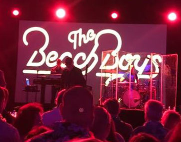
Beach Boys performance can’t distract ASEMV attendees

The attendees’ profound interest in exosomes was indicated on the opening evening of the meeting. At the outset, in outlining the logistics of the meeting, ASEMV president Dr. Gould explained that a late-breaking room change for Saturday night’s opening session had been occasioned by concern that the original room might be too noisy due to a performance taking place downstairs by The Beach Boys. The Beach Boys? Many thought that Dr. Gould was joking. But yes, the real Beach Boys were actually playing at a benefit event just downstairs from the ASEMV opening session, and yet, such was the audience’s interest in exosomes that no one moved. This reporter, however, could not resist checking out the iconic band after the opening ASEMV session had ended, and the photo here was taken of The Beach Boys who were indeed playing just downstairs. One of the original band members, Mike Love, was playing keyboard and singing. The Beach Boys performance was the highlight of a gala evening sponsored by Chimes (https://chimes.org/), a Baltimore-based international not-for-profit organization dedicated to assisting people with intellectual and behavioral challenges to achieve their fullest potential. It was an awesome backdrop to what would be an awesome ASEMV meeting.

Nearby International Human Virology meeting features major session on “Exosomes in health & disease”

And one further note is that the Institute for Human Virology (IHV), headed by legendary HIV virologist Dr. Robert Gallo, was holding its 20th International Meeting in the Four Seasons Hotel, right next to the Marriott where the ASEMV meeting was held.

Further indication of the exploding interest in exosomes was that the IHV meeting held a major session on exosomes this year. Titled “Exosomes in Health and Disease,” this session was listed second among nine sessions called out for special attention on the IHV meeting web page (https://www.ihv.org/ihvmeeting/). Areas of emphasis in this session ranged from cytokines in EVs to mechanisms of EVs in viral transmission.

Chairpersons of the IHV exosome session were Robert Gallo, MD, Director, Institute of Human Virology, University of Maryland School of Medicine, US, and Leonid Margolis, PhD, Senior Investigator, National Institute of Child Health and Human Development, US.

Speakers and topics included Xandra Breakefield, PhD, Professor of Neurology, Harvard Medical School / Genetist, Massachusetts General Hospital, “Extracellular Vesicle As Advance Forces in Cancer;” Dr. Margolis, “Not All Soluble Cytokines Are Soluble: Cytokines in Extracellular Vesicles Mediate Cell-Cell Communications;” Fatah Kashanchi, PhD, Former Director of Research, George Mason University, “Presence of HIV-1 RNA in Extracellular Vesicles from HIV-1 cART-Treated Cells;” Ayuko Hoshino, PhD, Instructor of Molecular Biology in Pediatrics, Weill Cornell Medical College, “Exosomal Protein Signatures: Mechanistic Insights and Biomarker Potential;” and Yoel Sadovsky, MD, Executive Director, Magee-Womens Research Institute, University of Pittsburgh, “Placental Exosomes in Maternal-Placental-Fetal Communication and Viral Resistance.”

Sponsors of ASEMV 2018 annual meeting

Sponsors of the ASEMV 2018 annual meeting included Particle Metrix, System Biosciences (SBI), iZON, Caris Life Sciences, nanoView Diagnostics, WAKO, ReNeuron, Norgen Biotek Corporation, Millipore Sigma, Beckman-Coulter, Wyatt Technology, AcouSort, Spectradyne, Fiber Cell Systems, ONI, abcam, Ceres Nano, Nanostics Precision Health, cellex, HansaBioMed Life Sciences, Lonza, and NanoTech.

Reference
Paget S. The distribution of secondary growths in cancer of the breast. The Lancet 133: 571-573. doi: 10.1016/S0140-6736(00)49915-0


Image: Kelsey Burke

This post originated as a press release from the University of Pennsylvania.

Cancerous tumors are more than a lump of cells growing out of control; they participate in active combat with the immune system for their own survival. Being able to evade the immune system is indeed a hallmark of cancer. Now, researchers from the University of Pennsylvania show that, to assist in the fight, cancer cells release biological “drones,” small vesicles called exosomes circulating in the blood and armed with the protein PD-L1, which causes T cells to tire before they have a chance to reach the tumor and do battle.

The work, published in the journal Nature (Chen et al., 2018), is a collaboration between Wei Guo of Penn’s School of Arts and Sciences and Xiaowei Xu of the Perelman School of Medicine. While primarily focused on metastatic melanoma, the team found that breast and lung cancer also release the PD-L1-carrying exosomes.

The research offers a paradigm-shifting picture of how cancers take a systemic approach to suppressing the immune system. In addition, it also points to a new way to predict which cancer patients will respond to certain checkpoint inhibitor drugs, which disrupt immune suppression to fight tumors, and a means of tracking the effectiveness of such therapies.

“Immunotherapies are life-saving for many patients with metastatic melanoma, but about 70 percent of these patients don’t respond,” says Guo, a professor of biology. “These treatments are costly and have toxic side effects, so it would be very helpful to know which patients are going to respond. Identifying a biomarker in the bloodstream could potentially help make early predictions about which patients will respond and, later on, could offer patients and their doctors a way to monitor how well their treatment is working.”

“Exosomes are tiny lipid-encapsulated vesicles with the diameter less than one-hundredth of a red blood cell. What we have found with these circulating exosomes is truly remarkable,” says Xu. “We collected blood samples from melanoma patients treated with anti-PD1 checkpoint inhibitor therapy. This type of liquid biopsy assay allows us to monitor tumor-related immune suppression with time.”

One of the most successful innovations in cancer therapy has been the use of checkpoint inhibitor drugs, which are designed to block attempts by cancer cells to suppress the immune system to allow tumors to thrive and spread. One of the primary targets for this class of drugs is PD-1, a protein on the surface of T cells. Tumor cells express PD-L1, which interacts with PD-1, effectively turning off that cell’s anti-cancer response. Blocking that interaction using checkpoint inhibitors reinvigorates T cells, allowing them to unleash their cancer-killing power on the tumor.

While it was known that cancer cells carried PD-L1 on their surface, Guo, Xu and colleagues found, in the new work, that exosomes from human melanoma cells also carried PD-L1 on their surface. Exosomal PD-L1 can directly bind to and inhibit T cell functions. Identifying the exosomal PD-L1 secreted by tumor cells provides a major update to the immune checkpoint mechanism, and offers novel insight into tumor immune evasion.

“Essentially exosomes secreted by melanoma cells are immunosuppressive.” Guo says. “We propose a model in which these exosomes act like drones to fight against T cells in circulation, even before the T cells get near to the tumor.”

Since a single tumor cell is able to secrete many copies of exosomes, the interaction between the PD-L1 exosomes and T cells provides a systemic and highly effective means to suppress anti-tumor immunity in the whole body. This may help explain why cancer patients have weakened immune systems.

Because exosomes circulate in the bloodstream, they present an accessible way of monitoring the cancer-T cell battle through a blood test, compared to a traditional, more-invasive tumor biopsy. After an acute phase of treatment, the researchers envision such a test as a way to monitor how well the drugs are keeping cancer cells in check.

By measuring pre-treatment levels of PD-L1, oncologists may be able to predict the extent of tumor burden in a patient and associate that with treatment outcome. In addition, a blood test could measure the effectiveness of a treatment. For example, levels of exosomal PD-L1 could indicate the level of T cell invigoration by immune checkpoint inhibitors.

“In the future, I think we will begin to think about cancers as a chronic disease, like diabetes,” says Guo. “And just as diabetes patients use glucometers to measure their sugar levels, it’s possible that monitoring PD-L1 and other biomarkers on the circulating exosomes could be a way for clinicians and cancer patients to keep tabs on treatment. It’s another step toward precision and personalized medicine.”


Reference

Chen G. et al. Exosomal PD-L1 contributes to immunosuppression and is associated with anti-PD-1 response. Nature (2018) 560: 382–386. doi: 10.1038/s41586-018-0392-8 PMID: 30089911


Guo and Xu coauthored the work with Penn’s Gang Chen, Alexander C. Huang, Wei Zhang, Min Wu, Jiegang Yang, Beike Wang, Honghong Sun, Wenqun Zhong, Bin Wu, Xiaoming Liu, Lei Guan, Tin Li, Shujing Liu, Ruifeng Yang, Youtao Lu, Liyun Dong, Suzanne McGettigan, Ravi Radhakrishnan, Junhyong Kim, Youhai H. Chen, Giorgos C. Karakousis, Tara C. Gangadhar, Lynn M. Schuchter, and E. John Wherry, as well as collaborators from Wuhan University, The Wistar Institute, Xi’an Jiaotong University, the University of Texas MD Anderson Cancer Center, and the Mayo Clinic.

The research was supported by the National Institutes of Health (GM111128, GM085146, AI105343, AI108545, AI082630, and AI117950), Parker Institute for Cancer Immunotherapy, American Heart Association, Tara Miller Melanoma Foundation, University of Pennsylvania, Wistar Institute, Dr. Miriam and Sheldon G. Adelson Medical Research Foundation, CAST Foundation, and NSFC Foundation.

Wei Guo is a professor of biology in the School of Arts and Sciences, and Xiaowei Xu is a professor of pathology and laboratory medicine and of dermatology in the Perelman School of Medicine at the University of Pennsylvania.

Wei Guo, Xiaowei Xu, and Gang Chen are listed as inventors on a patient owned by the University of Pennsylvania related to this work. Guo and Xu serve on the Scientific Advisory Board and have equities in Exo Bio, a company that has licensed the patent from the University of Pennsylvania.

This blog was adapted and simplified from a Spotlight in the Journal of Cell Biology.

Directed movement along a chemical gradient – chemotaxis – is a fundamental behavior of cells in the body during processes like inflammation, embryogenesis, and cancer metastasis. While the intracellular signaling mechanisms underlying chemotaxis have been investigated by many groups, it is not well known how stable gradients of chemoattractant chemicals are generated and maintained outside the cell, especially given the rapid diffusion of chemicals after secretion.

The amoeba Dictyostelium discoideum (fondly known as Dicty) is a model organism often studied since its biology can shed light on that of human cells. Recent work from Kriebel et al. (2018) showed that extracellular vesicles (EVs) are central to the biology of chemotaxis in Dicty by demonstrating that the main chemical Dicty follows during chemotaxis is synthesized within and released from EVs.

Dictyostelia are soil-living amoebas that undergo chemotaxis toward cyclic adenosine monophosphate (cAMP) under starvation conditions. As cAMP is released from the rear of migrating cells, chemotaxis toward cAMP leads to aggregation of the amoebas into multicellular structures.

Kriebel et al. previously showed (2008) that the enzyme that synthesizes cAMP, adenylyl cyclase (ACA), is present in multivesicular bodies (MVBs) located at the rear of migrating amoebas. They have now shown that leader cells release ACA-enzyme-containing vesicular trails and that follower cells stream onto them in a head-to-tail fashion. The vesicular trails are highly chemotactic and direct the migration of the follower cells.

Measurement of ATP, the precursor used by the enzyme to synthesize the product cAMP, revealed that it is also present inside EVs. Narrowing down from the 68 transporter proteins found in Dicty, the researchers identified the one protein – ABCC8 – that transports cAMP from the inside to the outside of EVs in order to promote chemotaxis. Dicty amoebas with that transporter knocked out exhibit much less chemotaxis and streaming behavior, and fluorescently tagged ABCC8 reintroduced into those amoebas is visible in the vesicular trails but not the plasma membrane.

Altogether, these data indicate that the entire machinery for generating and releasing chemoattractants is contained within EVs: the catalytically active enzyme (ACA), its substrate (ATP), the product (cAMP), and the transporter (ABCC8; see figure).

EVs secreted from Dictyostelia synthesize and release cAMP to promote chemotaxis. cAMP is converted from ATP by enzyme ACA in EVs and secreted through the ABCC8 transporter. Secreted cAMP makes a gradient and promotes chemotaxis of follower cells.

Overall, Kriebel et al. (2018) describe an elegant system by which chemotactic signals are generated and sustained to promote streaming in a complex multicellular system, and they elucidate a major mechanism by which cells leave a memory of themselves. EVs are left in a breadcrumb-like trail behind cells and continue sending chemotactic signals to surrounding cells. The finding that EVs act as cell-independent entities to not only carry but also generate bioactive products is important because it shows that they can amplify signals.

The same group made similar findings previously for neutrophils, one of the main sentry cells of the immune system (Majumdar et al., 2016). In both cases, the presence in EVs of enzymes that can generate a chemical product continuously amplifies and sustains a signal beyond that which would occur if the EVs carried just the chemical. The findings in amoeba and human cells together suggest that amplification of chemotactic signals is an important function of EVs that is conserved across organisms and necessary to promote effective directional sensing.

In a different context, it has been reported that precursor miRNAs can be processed into mature miRNAs in exosomes in a cell-independent manner due to the presence of the RNA interference–silencing complex (RISC) machinery in breast cancer–associated exosomes (Melo et al., 2014) and that this activity is important to promote tumor aggressiveness. Along with Melo et al. (2014), Kriebel et al. (2018) provide direct evidence that EVs can act as an independent machinery to regulate biological processes such as chemotaxis or tumorigenesis via enzymatic activities.

References

Kriebel PW, Barr VA, Rericha EC, Zhang G & Parent CA . (2008) Collective cell migration requires vesicular trafficking for chemoattractant delivery at the trailing edge. J. Cell Biol. 183:949–961. doi: 10.1083/jcb.200808105

Kriebel PW, et al. (2018) Extracellular vesicles direct migration by synthesizing and releasing chemotactic signals. J. Cell Biol. doi: 10.1083/jcb.201710170

Majumdar R, Tavakoli Tameh A & Parent CA. (2016) Exosomes mediate LTB4 release during neutrophil chemotaxis. PLoS Biol. 14:e1002336. doi: 10.1371/journal.pbio.1002336

Melo, et al. 2014. Cancer exosomes perform cell-independent microRNA biogenesis and promote tumorigenesis. Cancer Cell 26:707–721. doi: 10.1016/j.ccell.2014.09.005

Sung BH & Weaver AM. (2018) Directed migration: Cells navigate by extracellular vesicles. J. Cell Biol. doi: 10.1083/jcb.201806018

When someone finds out that I work at the National Institute on Aging, they usually ask me “How can I stop aging?”. Although aging is inevitable, it is well-known that individuals age at different rates and that certain groups age more rapidly than others. Hence, one of our laboratory’s objectives is to identify why certain population groups age differently, with the goal to find biomarkers that can tell us an individual’s biological age (how old you seem) versus chronological age (the actual time you have been alive).

Several years ago, we began this journey by examining whether small non-coding RNAs called microRNAs (miRNAs) change with age. We focused first on these regulatory RNAs because data has shown that these RNAs are particularly stable in biofluids, such as serum and plasma, and can be identified readily using small RNA sequencing. We identified several serum miRNAs that change significantly with age in both humans and rhesus monkeys (Noren Hooten, Fitzpatrick, et al. 2013). In most cases, their abundance in circulation decreases as we get older. Interestingly, some of these miRNAs target and negatively regulate the expression of various inflammatory markers, suggesting that decreased expression of these circulating miRNAs may contribute to higher levels of inflammatory markers that have already been observed in the elderly.

More recently we have focused on establishing a more complete extracellular RNA (exRNA) profile of human aging. To do so, we developed a sequencing pipeline that enables us to sequence both small and long RNAs in one sequencing reaction, which lowers both the cost and labor required (Dluzen, Noren Hooten, et al. 2018). Cataloging what is the “normal” distribution of exRNA in young and old individuals and identifying age-dependent differences will aid in establishing important references for the study of age-related disease. The Ensembl database classifies RNA into various categories, termed biotypes. We found that most RNA biotypes were similar in distribution between young and old, but several biotypes, including mitochondrial tRNAs (Mt_tRNA), mitochondrial ribosomal RNAs (Mt_rRNA), and unprocessed pseudogenes, were significantly higher in older individuals.


Figure 1. Changes in circulating factors with age. A decrease in circulating levels of specific miRNAs occurs with age. On the other hand, unprocessed pseudogenes, Mt_tRNAs, and Mt_rRNAs increase in abundance with age.

Pathway analysis revealed that RNAs related to mitochondria, response to oxidative stress, and chromatin remodeling were all enriched in the circulation of older individuals, providing potential clues as to what pathways may be deregulated as humans age. We also further validated our sequencing results in a larger cohort of individuals and found age-related changes in a messenger RNA, a small nucleolar RNA, a pseudogene transcript, a small nuclear pseudogene transcript, and several additional miRNAs.

What was very interesting was that we identified many circular RNAs (circRNAs) in serum, which we have named ex-circRNAs. Recent attention has been focused on circRNAs, as this class of ncRNAs may be important modulators of gene expression. CircRNAs have long half-lives (i.e. are stable) compared to mRNAs, making them an attractive new serum biomarker. However, little is known about ex-circRNAs, and I anticipate that this will be an active area of interest in the coming years.

As we have begun to establish an exRNA profile of human aging, there remain important unanswered questions in the field. Currently, we do not fully understand how exRNA in the circulation reflects the health status of our cells and tissues. It has also proven difficult to ascertain which cell type is contributing exRNA into the circulation. Further research is needed to better understand these questions.

Our identification of changes in circulating miRNAs and exRNAs establishes baseline references for how these biomarkers change with human age. As the risk for many diseases including cancer, heart disease, and neurological diseases increase with age, it is important to consider age when examining these factors in relation to a specific disease. Although we have not identified the “fountain of youth” or the “magic elixir” for aging, we hope that establishing these profiles with normal aging will soon help to identify circulating biomarkers that can distinguish individuals with faster biological aging that may result in shortened health span and life span.

References
Dluzen DF, Noren Hooten N, De S, Wood H, Zhang Y, Becker KG, Zonderman AB, Tanaka T, Ferrucci L & Evans MK. Extracellular RNA profiles with human age. Aging Cell 2018;e12785. PMID 29797538.

Noren Hooten N, Fitzpatrick M, Wood WH, De S, Ejiogu N, Zhang Y, Mattison JA, Becker JG, Zonderman AB & Evans MK. Age-related changes in microRNA levels in serum. Aging (2013) 5: 725-740. PMID 24088671.

This post originated as a press release from Linköping University.

The waste-management system of the cell appears to play an important role in the spread of Alzheimer’s disease in the brain. A new study, published in the prestigious scientific journal Acta Neuropathologica, has focused on small membrane-covered droplets known as exosomes. It was long believed that the main task of exosomes was to help the cell to get rid of waste products. In simple terms, they were thought of as the cell’s rubbish bags. However, our understanding of exosomes has increased, and we now know that cells throughout the body use exosomes to transmit information. It’s now known that the exosomes can contain both proteins and genetic material, which other cells can absorb.

The Linköping researchers have shown in the new study that exosomes can also transport toxic aggregates of the protein amyloid beta, and in this way spread the disease to new neurons. Aggregated amyloid beta is one of the main findings in the brains of patients with Alzheimer’s disease, the other being aggregates of the protein tau. As time passes, they form ever-increasing deposits in the brain, which coincides with the death of nerve cells. The cognitive functions of a person with Alzheimer’s disease gradually deteriorate as new parts of the brain are affected.

“The spread of the disease follows the way in which parts of the brain are anatomically connected. It seems reasonable to assume that the disease is spread through the connections in the brain, and there has long been speculation about how this spread takes place at the cellular level,” says Martin Hallbeck, associate professor in the Department of Clinical and Experimental Medicine at Linköping University and senior consultant of clinical pathology at Linköping University Hospital.

Cells became diseased
In a collaboration with researchers at Uppsala University, he and his co-workers have investigated exosomes in brain tissue from deceased persons. The research team at Linköping University found more amyloid beta in exosomes from brains affected by Alzheimer’s disease than in healthy controls. Furthermore, the researchers purified exosomes from the brains from people with Alzheimer’s disease, and investigated whether they could be absorbed by cells cultured in the laboratory.

“Interestingly, exosomes from patients were absorbed by cultured neurons, and subsequently passed on to new cells. The cells that absorbed exosomes that contained amyloid beta became diseased,” says Dr. Hallbeck.

The researchers treated the cultured neurons with various substances that prevent exosomes from being formed, released, or absorbed by other cells. They were able to reduce the spread of the aggregated amyloid beta between cells by disrupting the mechanism in these ways. The methods used in these laboratory experiments are not yet suitable for treating patients, but the discovery is important in principle.

“Our study demonstrates that it is possible to influence this pathway, and possibly develop drugs that could prevent the spreading. The findings also open up the possibility of diagnosing Alzheimer’s disease in new ways, by measuring the exosomes,” says Martin Hallbeck.

The research has received financial support from donors that include the Swedish Research Council, the Swedish Alzheimer’s Foundation, and the Swedish Brain Foundation.

Sinha MS, Ansell-Schultz A, Civitelli L, Hildesjö C, Larsson M, Lannfelt L, Ingelsson M & Hallbeck M. Alzheimer disease pathology propagation by exosomes containing toxic amyloid-beta oligomers. Acta Neuropathologica AOP 13 June 2018. doi: 10.1007/s00401-018-1868-1

Translation by George Farrants.

This commentary originally appeared as an Editor’s Choice in Science Translational Medicine. Thanks to STM and Steven Jay for permission to reprint here.

The sci-fi thriller I, Robot tells the story of robots attempting to take over the world based on their interpretation of the three governing laws of their programming. This plan is thwarted with the help of Sonny, a unique robot who can ignore the three laws due to being programmed differently. This movie illustrates how selective programming can be a powerful tool that can be used to turn a subset of a population against the rest. This same concept underlies the strategy of gene-directed enzyme prodrug therapy (GDEPT) for cancer, which involves specific delivery of a gene to cancer cells that allows for subsequent activation of a systemically administered prodrug into a toxic form only in cells where an enzyme encoded by the delivered gene is present. Several GDEPT strategies have advanced to clinical trials; however, the specificity and fidelity of gene delivery are still limiting factors to successful translation.

Toward addressing these limitations, Wang et al. describe the use of modified extracellular vesicles (EVs) for targeted delivery of mRNA to cancer cells overexpressing the HER2 receptor. EVs are nanoscale vesicles secreted by many cell types that have been co-opted for a variety of therapeutic applications. However, targeted delivery using EVs has been challenging, as has encapsulation of large nucleic acid cargo. To address cargo encapsulation, the authors applied a transfection-based approach to successfully load exogenous mRNA encoding for the enzyme HChrR6 into EVs. To address targeting, the authors created a novel chimeric protein consisting of a HER2 antibody fragment to target the receptor on cancer cells and the C1C2 domain of lactadherin, which interacts with the EV membrane. By mixing mRNA-loaded EVs with purified chimeric protein, the EVs were endowed with targeting capability for HER2-overexpressing cancer cells. Delivery of these EVs followed by systemic administration of the prodrug 6-chloro-9-nitro-5-oxo-5H-benzo-(a)-phenoxazine (CNOB) resulted in near complete growth arrest of orthotopically implanted HER2-overexpressing breast tumors in mice.

This report establishes a new and versatile approach for improving GDEPT that could be applied to a wide variety of cancers and other diseases. Significant barriers to translation of this approach remain, most notably the problem of scalability of EV-based approaches. However, the methods and strategy described are likely to have broad utility in further developing both GDEPT and therapeutic EVs.

Highlighted Article
J.-H. Wang, A. V. Forterre, J. Zhao, D. O. Frimannsson, A. Delcayre, T. J. Antes, B. Efron, S. S. Jeffrey, M. D. Pegram, A. C. Matin, Anti-HER2 scFv-directed extracellular vesicle-mediated mRNA-based gene delivery inhibits growth of HER2-positive human breast tumor xenografts by prodrug activation. Mol. Cancer Ther. (2018) 17:1133-1142. PMID:29483213 doi:10.1158/1535-7163.MCT-17-0827

Despite being one of the earliest known classes of non-coding RNA molecules, transfer RNAs (tRNAs) are still notoriously difficult to study. The challenge is largely due to this molecule’s secondary structure, chemical modifications to its constituent nucleotides (see figure), and the multiplicity of tRNA genes. As the number of non-coding RNA datasets proliferates, it is becoming increasingly important for tRNA genes to be accurately annotated. In a recent study, Thomas Tuschl from Rockefeller University and colleagues tackled this problem by developing a new protocol for sequencing tRNAs. The new method enabled them to assemble an atlas of human tRNAs for other researchers to use in analyzing their non-coding RNA data.

Hydro-tRNA Sequencing
Transfer RNAs have thermodynamically stable secondary and tertiary structures, and their constituent nucleotides are highly modified by RNA editing. Both of these characteristics are problematic for traditional RNA sequencing methods. The key to the Tuschl lab’s protocol, called hydro-tRNA sequencing (hydro-tRNAseq), is a partial alkaline hydrolysis step that breaks the 60-100 nucleotide-long tRNA into smaller fragments with fewer RNA modifications. These fragments, 19-35 nucleotides in size, have weaker secondary structure and fewer RNA modifications per fragment than the parent tRNA.

Applying the method to short RNA extracted from human embryonic kidney (HEK293) cells resulted in an increase in the fraction of reads mapped to tRNA between 2% and 40%, depending on the depth of sequencing. The short fragment length also improved read accuracy per base compared to standard tRNA sequencing.

To develop a thorough and representative reference set of human tRNAs, the HEK293 dataset was subjected to iterative cycles of mapping to existing reference tRNAs followed by manual curation. In each round, all transcripts with an error distance (number of mismatches, insertions, and deletions) of 1-2 from a given tRNA were kept as candidate reference sequences if they could be attributed to a tRNA isoacceptor (i.e. a different tRNA that binds to the same amino acid). If not, assuming that other mismatches were caused by misidentifying a modified base, transcripts with more than 10% mismatches compared to reference were expanded into a set of all possible combinations of RNA modifications and included in the reference pool (see figure). This mapping and selection process was repeated until there were no longer any modified positions left with a mismatch frequency over 10% compared to reference.

Candidate pre-tRNA genes were obtained by mapping the final tRNA reference sequences back to the genome. Altogether, this analysis was able to account for 93% of the 114 million reads in the deepest library of HEK293 cells’ tRNAs.

tRNA Modification Sites

tRNA Modification Sites
The team identified sites of modification from the high frequency of mismatches during mapping caused by read errors there during reverse transcription. Here the reference nucleotide is at ring center, known modification outside the ring, and frequency of each nucleotide read at that site inside the ring.
Source: Cell Reports

The Added Power of SSB PAR-CLIP
Though hydro-tRNAseq greatly improved the reference dataset of human tRNAs, there was still a risk that it alone would miss pre-tRNAs expressed at low levels or processed quickly into mature tRNA. Previous efforts to assay that ephemeral population employed ChIP-seq of POLR3, the polymerase that transcribes all tRNA genes, but doing so assumed that polymerase binding always led to expression and complete processing. The Tuschl lab focused instead on SSB, a protein that binds to the 3′ end of pre-tRNAs, immunoprecipitating tRNAs crosslinked to SSB using a method called PAR-CLIP. As predicted, almost half of the reads from their SSB PAR-CLIP experiments mapped to pre-tRNAs. Combining SSB PAR-CLIP with hydro-tRNAseq allowed the team to better identify mature and pre-tRNAs with improved, accurate, nucleotide-level resolution.

This study supplies the community with several new and useful resources. Hydro-tRNAseq provides a new method to overcome many of the struggles of tRNA sequencing analyses. Combining this method with SSB PAR-CLIP enabled the construction of a comprehensive atlas of pre-tRNAs and mature tRNAs in humans. This methodology can now be applied to study the tRNA complement in other species to further dissect tRNA biology.

Reference
Tasos Gogakos T, Brown M, Garzia A, Meyer C, Hafner M, & Tuschl T. Characterizing Expression and Processing of Precursor and Mature Human tRNAs by Hydro-tRNAseq and PAR-CLIP. Cell Reports (2017) 20: 1463-1475. doi: 10.1016/j.celrep.2017.07.029

In the Winter 2018 ERCC Newsletter, we review some of what was discussed at the ERCC9 conference last November. A major theme and continuing focus of research is finding the best methods for production of extracellular vesicles for therapeutic applications. We also highlight future directions in exRNA research and provide the schedule of upcoming ERCC web seminars. The next seminar is on Thursday, March 1st, at 2pm ET by Dr. David Wong of UCLA. He will discuss “Salivary exRNA and a New Horizon in Dental, Oral and Craniofacial Biology.” Dr. Wong and the ERCC are hosting symposia on the same topic at the annual meetings of the American and International Assocations for Dental Research, in Ft. Lauderdale, Florida in late March and London in July.

Please join us on the web and then in person!

You can download the newsletter here. Please be in touch at info@exRNA.org if you have an exRNA-related topic you would like us to cover in the Spring newsletter.

Our bodies are made up of trillions of cells that work together to keep us alive. A major challenge in their success is communication between cells in different parts of the body. Our cells have ingenious ways of overcoming this challenge, with exosomes emerging as key players. Exosomes are cell-derived vesicles that can carry cargo in the form of nucleic acids, lipids, or proteins from one cell to another. The sender cell packages cargo into an exosome, which then leaves the cell by being pinched off from the cell membrane. The exosome finds it way to a neighboring cell or into the bloodstream, from which it can be sent throughout the body. The exosome has signs on its surface that determine what cells can receive the cargo, so it only goes to the intended receiver. If a heart cell wants to talk to another heart cell, it puts markers on its exosomes that make them stick to other heart cells. When those exosomes are taken into the receiving cells, their cargo can bring about physiologic changes there.

Extracellular vesicles and their cargo. Source: BioProcess International.

Exosomes play a major role not only in our regular physiology but also in disease. One of the fields in which the role of exosomes is being uncovered is cardiovascular disease. For example, heart endothelial cells (cells that line the blood vessels) communicate with heart muscle cells via exosomes that contain microRNA, a kind of molecule that can decrease how many transcripts of a particular set of genes get made in the target cell. This process may play a role in the heart’s response to plaque formation. One can envision the possibility for engineering exosomes so that we can communicate with our bodies to treat or prevent disease. The lab of Dr. Susmita Sahoo at the Icahn School of Medicine at Mount Sinai is interested in doing just that.

Before talking about how Dr. Sahoo’s group is using exosomes in treating heart failure, let’s talk about a specific cause of heart failure: epitranscriptomics. You may or may not have heard of epigenetics, which is the study of heritable, chemical changes to DNA that do not change the sequence of the DNA. Epitranscriptomics is based on the exact same idea, but the change happens at the RNA level. One such epitranscriptomic modification is the addition or removal of methyl groups on adenosines within certain mRNAs in cells.

Structure of N6-Methyladenosine (m6A)

Heart muscle cells (cardiomyocytes) usually use electrical signals to interact and pulse in unison with a set rhythm. Work from Dr. Sahoo’s team suggests that decreasing levels of FTO, an enzyme that removes these methyl groups from RNA, leads to arrhythmia, a disturbance in that synchronized pulse. This finding is corroborated by the fact that failing hearts have low levels of FTO and elevated levels of mRNA methylation. Delivering exosomes with extra FTO to these cells might help them maintain healthy levels of FTO and decrease the chance of heart failure; this approach holds tremendous promise for the treatment of heart disease.

Dr. Sahoo’s research on exosomes is not limited to the failing heart. Recent work from her group suggests that a specific type of exosome, known to carry a marker called CD34 on its surface, improves angiogenesis, or formation of new blood vessels. Angiogenesis is a crucial step in healing after an injury. Dr. Sahoo’s group has shown that exosomes are able to improve healing in mice by providing microRNAs important for angiogenesis to cells near the site of injury. This work is not only important in helping patients after an injury, but it also teaches us about fundamental roles of microRNAs in angiogenesis and gene regulation.

We have outlined only some of the work going on in Dr. Sahoo’s lab. You can visit her website or watch her recent ERCC seminar to learn more about exosomes and her research on their role in cardiac medicine.