The American Association of Extracellular Vesicles Annual Meeting will be held in Houston, Texas from November 10th-13th, 2024.
Early bird registration ends September 1st!
For more information please visit 2024 Houston | AAEV
08.16.24
![]()
Common Fund Data Ecosystem (CFDE) Virtual Workshop:
“Biomarker discovery from extracellular (cell-free) RNA-seq and tissue of origin from GTEx RNA-seq profiles”
Thursday September 12th 2024
1-3 pm ET / 10am-Noon PT
Requires Free Registration by September 9th (below)
Join us for an engaging hands-on workshop where Srimeenakshi (Meenu) Srinivasan of the Louise Laurent Lab at the University of California-San Diego will demonstrate how to perform integrative data analysis utilizing data from two Common Fund Data Ecosystem (CFDE) Data Coordinating Center portals followed by an interactive Q&A session.
The featured use case will illustrate biomarker discovery from extracellular cell-free RNA-seq data with tissue of origin identification through integrative analysis of GTEx tissue-derived RNA-seq data and plasma-derived small RNA-seq data from the ERCC exRNA Atlas.
To conduct workshop exercises in real time, attendees wishing to take part in the hands-on analysis will need to have the following installed prior to dialing into the workshop as there will not be time to conduct installations during the workshop.
We also strongly encourage attendees to have Jupyter Notebook installed as well as this will be the platform used during the workshop. The Jupyter Notebook/files that will be used will be sent out via email.
Attendees who are not looking to conduct the analyses during the workshop are welcome to attend and observe.
A link to the virtual meeting will be provided in a confirmation email.
If you have any questions, please feel free to contact us at info@exrna.org
We look forward to seeing you there!
To learn more about the projects & resources that will be discussed please see:
Common Fund Data Ecosystem (CFDE): https://commonfund.nih.gov/dataecosystem
Extracellular RNA Communication Consortium (ERCC): https://exrna.org/
Genotype-Tissue Expression Project (GTEx): https://www.gtexportal.org/
![]()
Registration and more information:
When: October 17 – 19, 2024
Where: Bethesda North Marriott Hotel & Conference Center
Abstracts are due August 30, 2024
Registration deadline: September 25, 2024
The ASIC Annual Meeting is an intellectual “home” and support network for new and seasoned investigators, students, and postdocs. Over three days you’ll exchange ideas on emerging questions and cutting-edge developments in non-EV research including:
Extracellular Vesicles (EVs)
Extracellular Particles (EPs)
Particulate carriers of extracellular RNA (exRNA) as biological mediators, regulators, and diagnostic analytes.
The meeting also covers a broad range of intercellular communication processes including:
EVs
EPs
exRNA in Cancer
CNS Diseases
Infections (both bacterial and viral)
WHY ATTEND?
You’ll have a valuable opportunity to network with investigators from diverse scientific and clinical fields, discuss and advance the impact of EV/EP/ExRNA in diagnostics and treatments, and better understand the biogenesis of normal vs. disease states.
Are you new to the field? You can work with colleagues and mentors so you can improve your grants and develop their skills to become successful independent researchers.
Women and those from underrepresented groups are especially encouraged to attend and connect with seasoned researchers who have years of mentoring experience.
| This blog originated as a press release from the University of Houston. Thanks to them for allowing us to repost it here. |
Economical, Ultra-sensitive Biosensing in Point-of-Care Applications
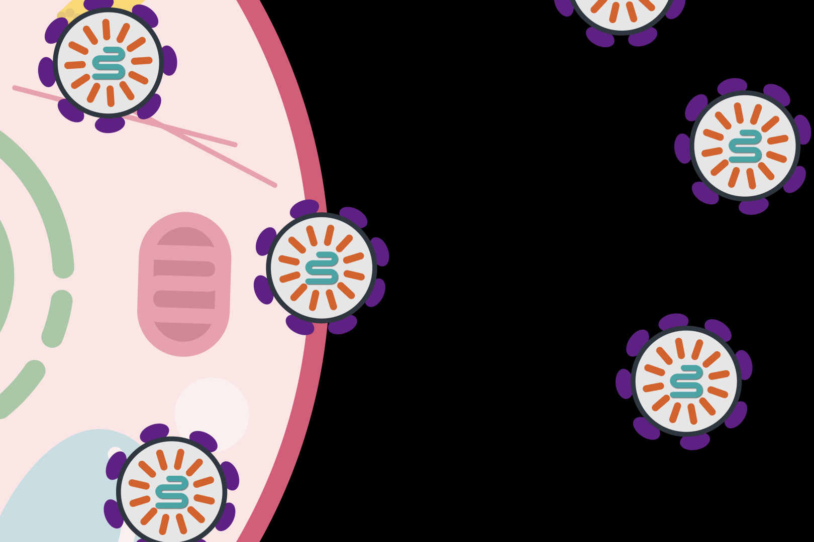
When it comes to cancer detection, size matters. Traditional diagnostic imaging cannot detect tumors smaller than a certain size, causing missed opportunities for early detection and treatment. Circulating tumor exosomes are especially small cancer biomarkers and easy to miss. These nanovesicles are composed of molecules that reflect the parental cells. But, because they are tiny (~30-150nm in diameter) and complex, the precise detection of exosome-carried biomarkers with molecular specificity has been elusive, until now.
Wei-Chuan Shih, professor of electrical and computer engineering at the University of Houston Cullen College of Engineering, reports the findings in IEEE Sensors Journal.
“This work demonstrates, for the first time, that the strong synergy of arrayed radiative coupling and substrate undercut can enable high-performance biosensing in the visible light spectrum where high-quality, low-cost silicon detectors are readily available for point-of-care application,” said Shih. “The result is a remarkable sensitivity improvement, with a refractive index sensitivity increase from 207 nm/RIU to 578 nm/RIU.”
| This blog originated as a press release from the Broad Institute of MIT and Harvard. Thanks to MIT News for allowing us to repost it here. |
Made of components found in the human body, the programmable system is a step toward safer, targeted delivery of gene editing and other molecular therapeutics.
Researchers from MIT, the McGovern Institute for Brain Research at MIT, the Howard Hughes Medical Institute, and the Broad Institute of MIT and Harvard have developed a new way to deliver molecular therapies to cells. The system, called SEND, can be programmed to encapsulate and deliver different RNA cargoes. SEND harnesses natural proteins in the body that form virus-like particles and bind RNA, and it may provoke less of an immune response than other delivery approaches.
| This blog originated as a press release from the Singopore-MIT Alliance for Research and Technology (SMART). Thanks to them for allowing us to repost it here. |
Four times faster than conventional PCR methods, a new approach called RADICA is highly specific, sensitive, and resistant to inhibitors.
● RApid DIgital Crispr Approach (RADICA) is a molecular rapid testing methodology that allows absolute quantification of viral nucleic acids in 40-60 minutes.
● RADICA is four times faster and significantly less expensive than conventional polymerase chain reaction (PCR) methods as it does not require costly equipment for precise temperature control and cycling.
● The method has been tested on SARS-CoV-2 synthetic DNA and RNA, Epstein–Barr virus in human B cells and serum, and can be easily adapted to detect other kinds of viruses.
Researchers from Critical Analytics for Manufacturing Personalized-Medicine (CAMP), an Interdisciplinary Research Group (IRG) at the Singapore-MIT Alliance for Research and Technology (SMART), MIT’s research enterprise in Singapore, have developed a new method for rapid and accurate detection of viral nucleic acids – a breakthrough that can be easily adapted to detect different DNA/RNA targets in viruses like the coronavirus.
This blog originated as a press release from The Ohio State University. Thanks to them for allowing us to repost it here.
Inserting genetic material into the body to treat diseases caused by gene mutations can work, scientists say – but getting those materials to the right place safely is tricky.
Scientists from The Ohio State University have reported in the journal Science Advances that the lipid-based nanoparticles they engineered, carrying two sets of protein-making instructions, showed in animal studies that they have the potential to function as therapies for two genetic disorders.
In one experiment, the payload-containing nanoparticles prompted the production of the missing clotting protein in mice that are models for hemophilia. In another test, the nanoparticles’ cargo reduced the activation level of a gene that, when overactive, interferes with clearance of cholesterol from the bloodstream.
 |
Dr. Yizhou Dong |
Each nanoparticle contained an applicable messenger RNA molecule that translates genetic information into functional proteins.
“We demonstrated two applications for lipid-like nanomaterials that effectively deliver their cargo, appropriately biodegrade, and are well-tolerated,” said Yizhou Dong, senior author of the study and associate professor of pharmaceutics and pharmacology at The Ohio State University.
“With this work, we have lowered potential side effects and toxicity and have broadened the therapeutic window. This gives us confidence to pursue studies in larger animal models and future clinical trials.”
This work builds upon a collection of lipid-like spherical compounds that Dong and colleagues had previously developed to deliver messenger RNA. This line of particles was designed to target disorders involving genes that are expressed in the liver.
The team experimented with various structural changes to those particles, effectively adding “tails” of different types of molecules to them, before landing on the structure that made the materials the most stable. The tiny compounds have a big job to do: embarking on a journey through the bloodstream, carrying molecules to the target location, releasing the ideal concentration of messenger RNA cargo at precisely the right time, and safely degrading.
The tests in mice suggested these particles could do just that.
The researchers injected nanoparticles containing messenger RNA holding the instructions to produce a protein called human factor VIII into the bloodstream of normal mice and mouse models for hemophilia. A deficiency of this protein, which enables blood to clot, causes the bleeding disorder. Within 12 hours, the deficient mice produced enough human factor VIII to reach 90 percent of normal activity. A check of the organs of both protein-deficient mice and normal mice showed that the treatment caused no organ damage.
“It can be helpful to think of this as a protein-replacement therapy,” Dong said.
In the second experiment, nanomaterials were loaded with two types of instructions: messenger RNA carrying the genetic code for a DNA base editor, and a guide RNA to make sure the edits occurred in a specific gene in the liver called PCSK9. Dozens of mutations that increase this gene’s activity are known to cause high cholesterol by reducing clearance of cholesterol from the bloodstream.
Analyses showed that the treatment resulted in the intended mutation of about 60 percent of the target base pairs in the PCSK9 gene, and determined that only a low dose was needed to produce high editing effect.
Dong credited academic and industry partners for helping advance this work. Co-corresponding authors include Denise Sabatino of Children’s Hospital of Philadelphia and Delai Chen from Boston-based Beam Therapeutics, who provided expertise in hemophilia and DNA base editing, respectively.
Dong and first author Xinfu Zhang are inventors on patent applications filed by Ohio State related to the lipid-like nanoparticles. This technology has been licensed for further clinical development.
This work was supported by the National Institute of General Medical Sciences, the National Heart, Lung and Blood Institute, and a startup fund from Ohio State’s College of Pharmacy.
Additional co-authors are Giang N. Nguyen of Children’s Hospital of Philadelphia; Weiyu Zhao, Chengxiang Zhang, Chunxi Zeng, Jingyue Yan, Shi Du, Xucheng Hou, Wenqing Li, Justin Jiang, Binbin Deng and David McComb of Ohio State; and Robert Dorkin, Aalok Shah, Luis Barrera, Francine Gregoire and Manmohan Singh of Beam Therapeutics.
Reference
Zhang X et al. Functionalized lipid-like nanoparticles for in vivo mRNA delivery and base editing. (2020) Sci Adv 6:eabc2315. doi: 10.1126/sciadv.abc2315. PMID: 32937374.
11.23.20
The ASEMV2020 organizing committee would like to congratulate the winners of this year’s Young Investigator Awards. There were three speaker awards, for talks by a Young Investigator, a postdoctoral scholar, and a Ph.D. candidate. There are also two poster winners.
Moran Amit
Assistant Professor
Department of Head and Neck Surgery – Research
Division of Surgery
University of Texas MD Anderson Cancer Center
for work on the role of p53 and axonogenesis in cancer
Frederik Verweij
Post-Doctoral Fellow
Team van Niel
Institute of Psychiatry and Neuroscience of Paris
for research on EV biology in a zebrafish model system
See Dr. Verweij’s recent #WebEVTalk outlining the zebrafish model system for tracking EVs.
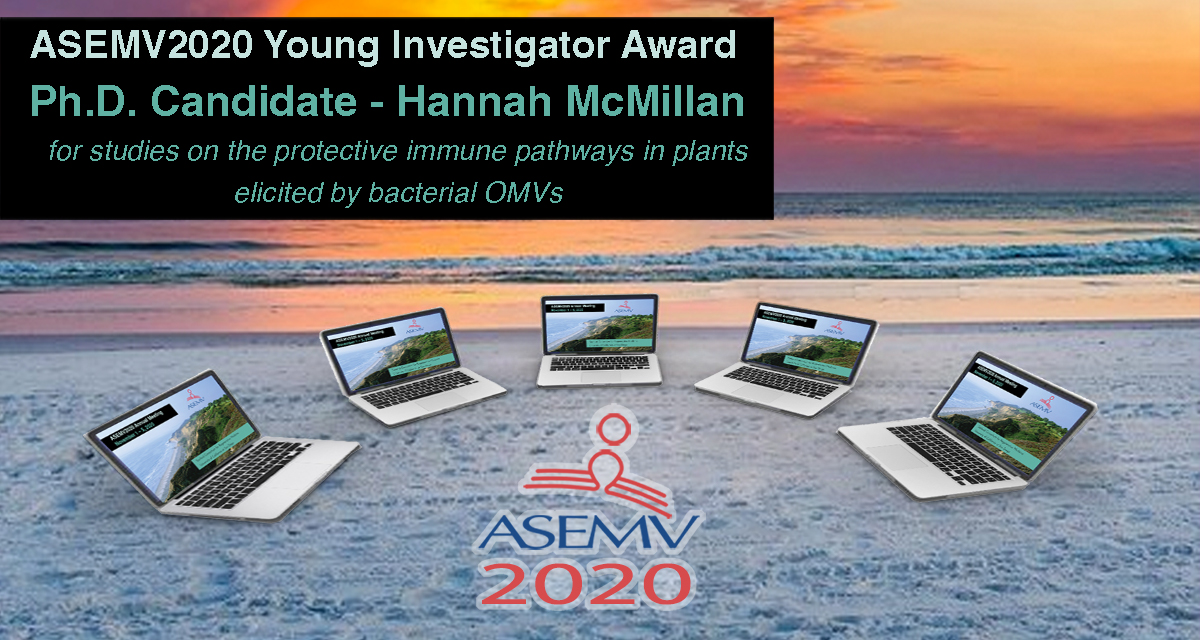
Hannah McMillan
Ph.D. Candidate
Kuehn Lab
Department of Molecular Genetics and Microbiology
Duke University
for studies on the protective immune pathways in plants elicited by bacterial OMVs

Killian O’Brien
Post-Doctoral Fellow
Breakefield Lab
Harvard Medical School &
Massachusetts General Hospital
for research on understanding the intracellular fate of EV-delivered content
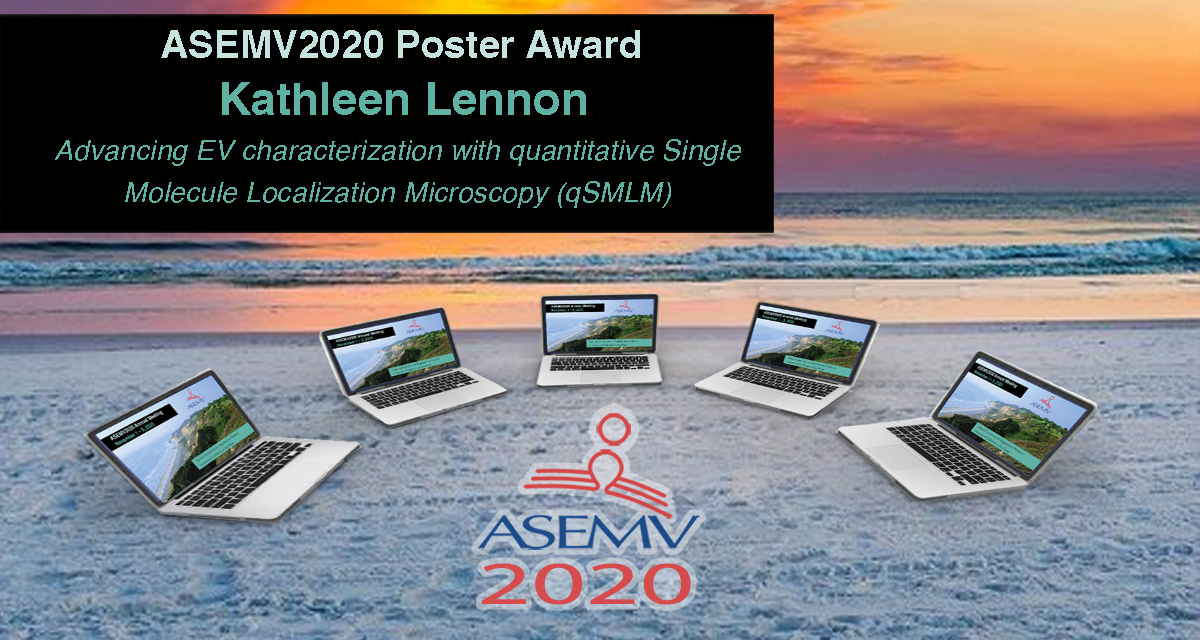 Kathleen Lennon
Ph.D. Candidate
Talisman Lab
Irell and Manella Graduate School of Biological Sciences
City of Hope
for work on EV characterization using quantitative Single Molecule Localization Microscopy (qSMLM)
| This blog originated as a press release from Duke Health. Thanks to them for allowing us to repost it here. |
DURHAM, N.C. – A team of Duke Health scientists have identified biomarkers that accurately identify numerous viral infections across the clinical stages of disease, advancing a potential new way to guide treatment, quarantine decisions, and other clinical and public health interventions in the setting of endemic and pandemic infectious diseases.
The blood-based test uses a gene expression assay to correctly predict nine different respiratory viral infections including influenza, enterovirus, adenovirus, and coronaviruses known to cause common colds. It shows the body’s genes responding to a pathogen before symptoms are present.
This blog originated as a press release from the University of Sussex. Thanks to them for allowing us to repost it here.
Scientists at the University of Sussex have identified a potential pattern within blood which signals the presence of motor neuron disease; a discovery which could significantly improve diagnosis.
Currently, it can take up to a year for a patient to be diagnosed with amyotrophic lateral sclerosis (ALS), more commonly known as motor neuron disease (MND).
But after comparing blood samples from patients with ALS, those with other motor-related neurological diseases, and healthy patients, researchers were able to identify specific biomarkers which act as a diagnostic signature for the disease.