Other

The National Institute of Diabetes and Digestive and Kidney Diseases (NIDDK) has announced a new funding opportunity for characterization of islet-derived extracellular vesicles (EVs) for improved detection, monitoring, classification, and treatment of Type 1 Diabetes (T1D).

This initiative will support the development of tools and experimental platforms for the purification and characterization of EVs originating from the human pancreatic islet and its broader tissue environment in healthy individuals, and individuals with T1D or at-risk of developing the disease. It will also support the exploration of the contribution of pancreatic EV biology to islet function, dysfunction and T1D disease initiation; the development of EV-based diagnostic tools for disease monitoring and classification; and the use of pancreatic EV biology to identify novel therapeutic targets.

A letter of intent to apply for the grant must be sent by October 3, 2021.

For more information, see https://grants.nih.gov/grants/guide/rfa-files/rfa-dk-21-016.html.

Extracellular vesicles (EVs) regulate many processes in the healthy body. They also play a role in cancer, sending signals between cells in the tumor microenvironment. EVs can stimulate tumor cell migration, invasion, blood vessel growth, immune response, and cell survival, as well as metastasis. However, we know little about the cargo of these EVs that play such diverse roles. Analysis of vesicle cargo can shed light on the molecular mechanisms of vesicle biology and be helpful in disease diagnosis and prognosis.

I am lucky to be a member in Jan Lötvall’s lab in Gothenburg, Sweden, which pioneered the field of extracellular vesicles with the early discovery of exosomes shuttling RNA between cells. An exciting collaboration with Yong Song Gho from POSTECH in South Korea led us to develop a new approach to isolate vesicles from human tumor tissues. Using this technology, we were able to isolate and characterize subpopulations of extracellular vesicles from melanoma metastatic tissue. We just published our findings in the Journal of Extracellular Vesicles. Jan Lötvall also discussed them in a recent ERCC webinar.

Continue reading

This blog originated as a press release from the University of Sussex.

New research by scientists at the University of Sussex could be the first step towards developing a blood test to diagnose the most aggressive type of brain tumour, known as Glioblastoma.

A team from Professor Georgios Giamas’ lab at the University of Sussex has identified novel biomarkers within bodily fluids, which signal the presence of the tumour. Dr. Giamas is Professor of Cancer Cell Signalling in the School of Life Sciences.

Cancer biomarkers are molecules that are either exclusively found or over-expressed in cancer cells, as compared to ‘normal’, healthy cells. Biomarkers can be considered as biological signatures for a disease, as they indicate the presence of cancer in the body.

In a new paper published in the Nature journal Communications Biology, Professor Giamas and his team describe particular biomarkers that are associated with extracellular vesicles – small ’packages’ released by cells into bodily fluids so cells can communicate with each other.

Continue reading

This blog post originated as a press release from Vanderbilt University Medical Center.

A report by researchers at Vanderbilt University Medical Center has shattered conventional wisdom about how cells, including cancer cells, shed DNA into the bloodstream: they don’t do it by packaging the genetic material in tiny vesicles called exosomes.

Their findings, reported April 4 in the journal Cell, have important implications for the development of “liquid biopsies” that could speed cancer diagnosis and improve treatment by detecting tumor-specific genetic material in the blood.

“It’s been a big deal that there is supposedly DNA in exosomes … (and) you could isolate these exosomes in a relatively simple way,” said the paper’s first author, Dennis Jeppesen, PhD. The problem is exosomes don’t contain DNA.

“Exosomes are not your target,” said Jeppesen, a research fellow in the lab of Robert Coffey, Jr., MD, who is internationally known for his studies of colorectal cancer. Instead, Jeppesen and his colleagues propose a new model for how DNA is actively secreted by cells.

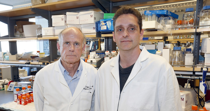
Research by Robert Coffey, MD, left, Dennis Jeppesen, PhD, and colleagues has revealed a new way cells shed DNA into the bloodstream.
(Photo by Steve Green / Vanderbilt University)

Greater precision in determining how DNA, RNA, and proteins are packaged and secreted from cells “is crucial for identification of biomarkers and design of future drug interventions,” the researchers concluded.

Coffey, Ingram Professor of Cancer Research in the Vanderbilt University School of Medicine, predicted the paper will “set the field on a firmer foundation” to understand what’s in exosomes and what’s not, and how exosomes might be used as biomarkers or therapeutic targets.

Coffey’s team is part a nationwide consortium funded by the National Institutes of Health (NIH) to study the role of extracellular RNA in diseases including diabetes, glaucoma, muscular dystrophy, and cancer.

Last month the NIH announced the publication of what it called a “landmark collection” of scientific papers in the Cell family of journals on the biology and possible clinical applications of extracellular RNA. Two of those papers, including the paper about exosomes, came from Coffey’s lab.

Virtually every cell in the body releases DNA, RNA, proteins, lipids, and other particles. These so-called “nanoparticles” are thought to be a way that cells communicate with each other. But they also might signal cancer to spread — or metastasize — to other parts of the body.

Cancer by its very nature is constantly evolving. That trait enables many tumors to escape or to become resistant to chemotherapy and other efforts to destroy them.

The genetic material released by cancer cells, however, may also reveal their points of vulnerability. A simple blood test that picks up these circulating cancer clues therefore could lead to earlier diagnoses and more effective treatments.

The problem is how to snag cancer-specific genetic material from the sea of circulating nanoparticles. Each milliliter of blood (it takes 5 milliliters to fill a teaspoon) contains a quadrillion (a thousand trillion) nanoparticles, and at least 100 million small extracellular vesicles.

Many of these vesicles are exosomes, which are known to carry extracellular RNA. Exosomes can be identified by the proteins (called antigens) that sprout from their surfaces. Identifying specific exosomes is one thing; determining what they carry is quite another.

The traditional way is to use high-speed centrifugation to spin exosomes out of a blood sample and into a “pellet” at the bottom of a test tube. Biochemical methods are then used to characterize the RNA, DNA, and proteins in the pellet.

The assumption was that the pellet contained only exosomes and their cargoes. But Jeppesen, who earned his PhD in Molecular Medicine at Aarhus University in Denmark, was skeptical. He decided to take a different approach.

Using a technique called high-resolution density gradient fractionation, Jeppesen and his colleagues separated blood components based on their buoyant density. They found that exosomes floated at a relatively low density while higher-density proteins, including those that bind DNA, sank to a lower gradient.

“We showed this for multiple cancer cell lines,” he said. “We also see the same kind of thing in normal cells.”

To isolate and study exosomes apart from the traditional pellet, Jeppesen and his colleagues developed another technique they called direct immunoaffinity capture. They coated magnetic beads with a “capture antibody” that targeted one of the known proteins, or antigens, on the exosome surface.

That’s how they were able to determine that exosomes don’t carry DNA.

The DNA found in pellets must be secreted by the cell in other ways. One way, the Vanderbilt researchers reported, is through the formation of novel hybrid organelles termed amphisomes.

“We can actually see these amphisomes traffic to the cell surface,” Jeppesen said. “Now it’s possible to say with greater precision what’s in the exosome and what’s in these other vesicles. Now you have an idea of what is the target you’re looking for.”

In the Winter 2018 ERCC Newsletter, we review some of what was discussed at the ERCC9 conference last November. A major theme and continuing focus of research is finding the best methods for production of extracellular vesicles for therapeutic applications. We also highlight future directions in exRNA research and provide the schedule of upcoming ERCC web seminars. The next seminar is on Thursday, March 1st, at 2pm ET by Dr. David Wong of UCLA. He will discuss “Salivary exRNA and a New Horizon in Dental, Oral and Craniofacial Biology.” Dr. Wong and the ERCC are hosting symposia on the same topic at the annual meetings of the American and International Assocations for Dental Research, in Ft. Lauderdale, Florida in late March and London in July.

Please join us on the web and then in person!

You can download the newsletter here. Please be in touch at info@exRNA.org if you have an exRNA-related topic you would like us to cover in the Spring newsletter.

We are pleased to announce the publication of miRandola 2017 in Nucleic Acids Research, Database issue 2018!
Citation:
miRandola 2017: a curated knowledge base of non-invasive biomarkers.
Francesco Russo*, Sebastiano Di Bella, Federica Vannini, Gabriele Berti, Flavia Scoyni, Helen V. Cook, Alberto Santos, Giovanni Nigita, Vincenzo Bonnici, Alessandro Laganà, Filippo Geraci, Alfredo Pulvirenti, Rosalba Giugno, Federico De Masi, Kirstine Belling, Lars J. Jensen, Søren Brunak, Marco Pellegrini, Alfredo Ferro.

*Correspondence to francesco.russo@cpr.ku.dk
URL: Nucleic Acids Research
Website: https://mirandola.iit.cnr.it/

Thanks to Laurence de Nijs and the European College of Neuropsychopharmacology (ENCP) for allowing us to adapt their press release into a blog.


Individuals affected with PTSD (Post-Traumatic Stress Disorder) demonstrate changes in microRNA (miRNA) molecules associated with gene regulation. A controlled study, involving Dutch military personnel on deployment to a combat zone in Afghanistan, provided evidence for the role of blood-based miRNAs as candidate biomarkers for symptoms of PTSD. This finding may offer an approach towards screening for symptoms of PTSD, and it holds promise for understanding other trauma-related psychiatric disorders. Limitations of the study are that this was a small pilot study, and the findings need to be validated, extended, and confirmed. First results were presented at the 30th conference of the European College of Neuropsychopharmacology (ENCP) in Paris in early September.

PTSD is a psychiatric disorder which can manifest following exposure to a traumatic event, such as combat, assault, or natural disaster. Among individuals exposed to traumatic events, only a minority of individuals will develop PTSD, while others will show resiliency. Little is known of the mechanisms behind these different responses. The last few years have seen much attention given to whether the modification and expression of genes – epigenetic modifications – might be involved. But there are several practical and ethical challenges in designing a research study on humans undergoing such experiences, meaning that designing relevant study approaches is difficult.

A research group from the Netherlands worked with just over 1,000 Dutch soldiers and the Dutch Ministry of Defense to study changes in biology in relation to changes in presentations of symptoms of PTSD in soldiers who were deployed to a combat zone in Afghanistan. In a longitudinal study, they collected blood samples before deployment as well as 6 months after deployment. Most of the soldiers had been exposed to trauma, and some of the soldiers had developed symptoms of PTSD.

For this pilot study, from the initial group, 24 subjects were selected in 3 subgroups of 8. Eight of the soldiers had developed symptoms of PTSD; 8 had endorsed traumatic experiences but had not developed symptoms of PTSD; and another 8 had not been in serious traumatic circumstances and served as a control group. Using modern sequencing techniques, several types of miRNAs for which the blood levels differed between the groups were identified.

MiRNAs (Micro RiboNucleic Acids) are small molecules with chemical building blocks similar to DNA. Unlike the more famous DNA, miRNAs are typically very short – comprising only around 20 to 25 base units (the building blocks of nucleic acids), and they do not code, in other words they do not specify the production of a protein or peptide. However, they have very important roles in biology (every miRNA regulates the expression, and thereby also the activity of several other genes), and they are known to regulate the impact of environmental factors on biology. In addition, brain-derived miRNA can circulate throughout the human body and can be detected in the blood.

Differences in miRNA levels have been associated with certain diseases, such as some cancers, kidney disease, and even alcoholism. This regulatory role makes them also a candidate for investigation in PTSD.

“We discovered that these small molecules, called miRNAs, are present in different amounts in the blood of persons suffering from PTSD compared to trauma-exposed and control subjects without PTSD,” said first author Dr Laurence de Nijs of Maastricht University.

“We identified over 900 different types of these small molecules. 40 of them were regulated differently in people who developed PTSD, whereas there were differences in 27 of the miRNAs in trauma-exposed individuals who did not develop PTSD.”

“Interestingly, previous studies have found circulating miRNA levels to be not only correlated with different types of cancer, but also with certain psychiatric disorders including major depressive disorders. These preliminary results of our pilot study suggest that miRNAs might indeed be candidates as predictive blood markers (biomarkers) to distinguish between persons at high and low risk of developing PTSD. However, several steps need to be performed before such results can really have an impact on the larger field and in clinical practice. In addition to working towards biomarkers, the results may also provide novel information about the biological mechanisms underlying the development of PTSD.”

Dr de Nijs explained:
“Most of our stressful experiences don’t leave a long-lasting psychological scar. However, for some people who experience chronic severe stress or really terrible traumatic events, the stress does not go away. They are stuck with it, and the body’s stress response is stuck in ‘on’ mode. This can lead to the development of mental illness such as PTSD.

These individuals experience symptoms including re-experiencing of the traumatic event through flashbacks or recurrent nightmares, constant avoidance of reminders of the event, negative mood, and extreme arousal. This can manifest itself through insomnia and or hyper-alertness. Individuals with PTSD are six times more at risk of committing suicide and having marital problems, and the annual loss of productivity is estimated to be approximately $3 billion. Currently, there is no definite cure for patients with PTSD, and available treatments often are not effective.”

Commenting, Professor Josef Zohar (Ex-ECNP Chair, Tel Aviv, Israel) said:
“The relevance of a better understanding of stress-related events is unfortunately becoming clearer and clearer after each terror attack. This work points to an innovative avenue regarding the potential identification of risk factors for susceptibility to developing post-traumatic stress disorder.”


Funding: Dr de Nijs was awarded a Marie Curie fellowship grant by the European Union to perform this study, within a network of other expert scientists in PTSD and epigenetics. The Dutch cohort of soldiers (PRISMO) was funded through the Dutch Ministry of Defence.

Extracellular vesicles, such as exosomes and microvesicles, are small vesicular particles that are constantly being produced and shed by cells. Due to their natural origin, and their ability to efficiently deliver their cargo to target cells and alter biological functions, exosomes attracted researchers to study their potential use as drug delivery systems. In the past few years, numerous studies have reported the effective use of exosomes to deliver therapeutic cargo ranging from miRNA, siRNA and even small molecule drugs in both in vitro cell models and in vivo animal models. However, since exosomes are cell-derived vesicles, it is unclear how these natural carriers of biomolecules may induce immune responses or induce toxicity either in animal models of disease or eventually in humans as we progress toward clinical evaluation of exosomes in healthy volunteers or in patients. Furthermore, what can we conclude about the presence or lack of immunogenicity or toxicity in our animal models as we work toward delivery of exosomes in humans?

Our lab has been studying the production of therapeutic exosomes using genetically engineered HEK293T cells for treatment of hepatocellular carcinoma (HCC). We developed engineered HEK293T cells that endogenously package miR-199a-3p, a miR commonly downregulated in HCC, into exosomes, and we are evaluating these and also exosomes exogenously loaded with therapeutic miRs in vitro and in vivo. Although demonstrating in vivo efficacy is a major milestone for all drug development efforts, understanding the potential toxicities and immunogenic responses associated with exosome therapy is equally important. The ability to identify and characterize adverse responses in preclinical models is critical to the drug development process and a necessary component of an Investigational New Drug (IND) application. Therefore, approaches to characterizing potential toxicities and immune responses induced by exosomes will be a necessary component of any effort to develop therapeutic exosomes.

In our article, “Comprehensive toxicity and immunogenicity studies reveal minimal effects in mice following sustained dosing of extracellular vesicles derived from HEK293T cells,” that was just published online in the Journal of Extracellular Vesicles, we provide a general template process for comprehensively evaluating toxicity and immunogenicity of therapeutic exosomes in preclinical animal models. We started by dosing mice with wild type or engineered HEK293T-derived exosomes over a period of three weeks. Mice received 10 doses via intraperitoneal and intravenous routes of injection, and blood samples were collected at various times throughout the 3-week study. Animals were euthanized 24 hours after the last dose, and blood and all organs were collected from each animal for gross necropsy and evaluation of various markers of immune response and potential exosome-induced toxicity markers.

This study demonstrates one approach to immunogenicity and toxicity evaluations of human-derived exosomes in mice, and it highlights some of the variables that must be considered during these evaluations. For example, what are appropriate animal models in which to study immunogenicity and toxicity? What doses and dose regimens should be evaluated? How might the cell type from which the exosomes were harvested impact immunogenicity and toxicity? Just as with efficacy evaluations in animals, comprehensive study of these other factors will be necessary for safely moving therapeutic exosomes into human trials.

The microRNA miR-155 plays a significant role in physiological and pathological processes in humans by blocking the functions of functionally important messenger RNAs of protein coding genes. We found that miR-155 was present in higher levels in cancers resistant to chemotherapy. By studying the association of miR-155 and tumor suppressor TP53 with cancer survival in 956 patients with lung cancer, chronic lymphocytic leukemia and acute lymphoblastic leukemia, we demonstrated that miR-155 induces resistance to multiple chemotherapeutic agents in vitro, and that blocking or down-regulating miR-155 successfully resensitizes tumors to chemotherapy in vivo. We found that high levels of miR-155 and low levels of TP53 characterize the tumors from lung cancer patients with shorter survival time. Our findings support the existence of a miR-155/TP53 feedback loop involved in resistance to chemotherapy. To target this feedback loop and effectively alter resistance to therapy, we have developed a therapeutic nanoformulation of anti-miR-155 in a lipid nanoparticle (DOPC) and have shown it to be non-toxic in vivo for further pre-clinical work.

We thank our co-authors for their work and discussions that led to this blog.

miR-155 TP53 and resistance to chemotherapy

Cancer cells actively reprogram gene expression to promote their ability to produce tumors. One way this reprogramming is carried out is by subverting the main routes of cell-to-cell communication by loading exosomes (vesicles that bud off from cells) with specific miRNAs that either promote or suppress tumors and then releasing them into the tumor microenvironment. In a Cancer Research paper published online recently (Kanlikilicer et al., 2016), we found that miR-6126, a miRNA that was reported to be correlated with better overall survival in high-grade serous ovarian cancer patients, is ubiquitously removed from ovarian cancer cells via exosomes.

miR-6126 is ubiquitously removed from ovarian cancer cells via exosomes

We found that miR-6126 suppresses tumors by directly targeting integrin ß1, a key regulator of cancer cell metastasis. Treatment of orthotopic mouse models of ovarian cancer with miR-6126 reduced tumor growth, proliferating cells, and microvessel density. Our findings provide new insights into the role of exosomes in mediating tumor progression and suggest a new therapeutic approach to disrupt the origin and growth of tumors.

Citation:
Ubiquitous release of exosomal tumor suppressor miR-6126 from ovarian cancer cells
Pinar Kanlikilicer, Mohammed Saber, Recep Bayraktar, Rahul Mitra, Cristina Ivan, Burcu Aslan, Xinna Zhang, Justyna Filant, Andreia M Silva, Cristian Rodriguez-Aguayo, Emine Bayraktar, Martin Pichler, Bulent Ozpolat, George A Calin, Anil K. Sood and Gabriel Lopez-Berestein*
Cancer Res. October 14, 2016 doi: 10.1158/0008-5472.CAN-16-0714
*Corresponding author