Extracellular vesicles (EVs) regulate many processes in the healthy body. They also play a role in cancer, sending signals between cells in the tumor microenvironment. EVs can stimulate tumor cell migration, invasion, blood vessel growth, immune response, and cell survival, as well as metastasis. However, we know little about the cargo of these EVs that play such diverse roles. Analysis of vesicle cargo can shed light on the molecular mechanisms of vesicle biology and be helpful in disease diagnosis and prognosis.
I am lucky to be a member in Jan Lötvall’s lab in Gothenburg, Sweden, which pioneered the field of extracellular vesicles with the early discovery of exosomes shuttling RNA between cells. An exciting collaboration with Yong Song Gho from POSTECH in South Korea led us to develop a new approach to isolate vesicles from human tumor tissues. Using this technology, we were able to isolate and characterize subpopulations of extracellular vesicles from melanoma metastatic tissue. We just published our findings in the Journal of Extracellular Vesicles. Jan Lötvall also discussed them in a recent ERCC webinar.
Our first challenge was to find vesicles in metastatic melanoma tumor tissues. Using transmission electron microscopy, we showed that the tumor microenvironment is a complex world composed of different types of cells and structures with vesicles present between them.
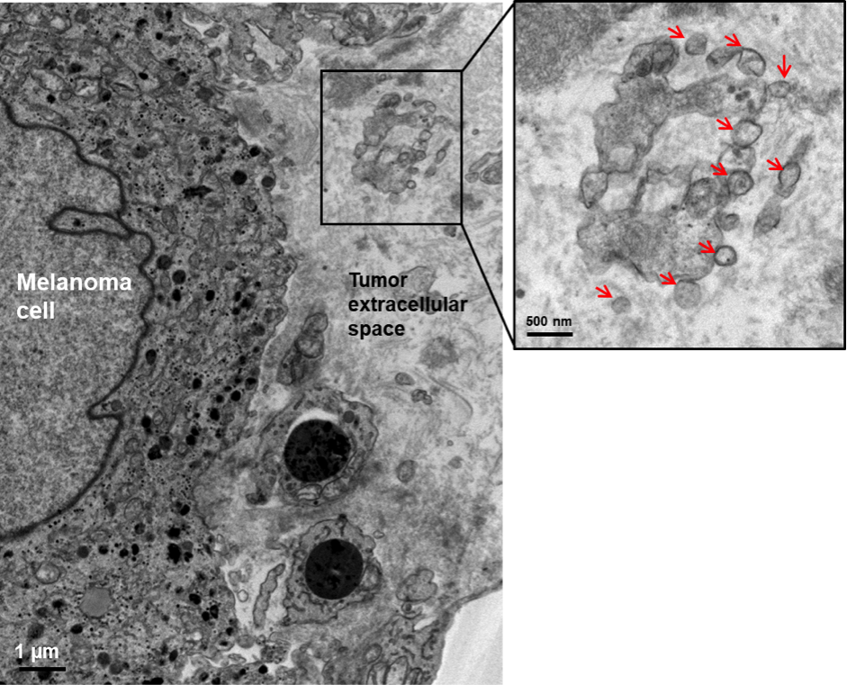
Transmission electron micrograph of melanoma metastatic tissue showing a large tumor cell and two lymphocytes. Black stain, possibly melanin, is clearly visible inside the melanoma cells, which are recognizable from their characteristic cell membrane. The higher magnification image shows vesicles (red arrows) in the extracellular space.
In this study, we performed a detailed proteomics analysis of EVs isolated from metastatic melanoma tissues from 27 patients. We identified numerous new EV proteins, including potential biomarkers for metastatic melanoma.
Studying extracellular vesicles in tumor tissues is important, because, compared to cell lines, tumor tissues more closely approximate the situation in vivo. EVs from tumor tissue are more likely to represent the full array of vesicle behaviors and populations in the tumor microenvironment. Furthermore, to develop a non-invasive test for cancer, we must use biofluids such as circulating plasma, where vesicles from all over the body intermingle. A proteomic snapshot of vesicles isolated directly from tumor tissue can help target the search for disease-specific biomarker in that complex mixture. We trust the tools and experiments developed in this work will contribute to our field’s understanding of EV function in complicated tissues such as the metastatic melanoma tumor.
Reference
Crescitelli R, Lässer C, Jang SC, Cvjetkovic A, Malmhäll C, Karimi N, Höög J.L, Johansson I, Fuchs J, Thorsell A, Gho YS, R, Olofsson Bagge R, Lötvall J Subpopulations of extracellular vesicles from human metastatic melanoma tissue identified by quantitative proteomics after optimized isolation. Journal of Extracellular Vesicles 9:1, 1722433 doi: 10.1080/20013078.2020.1722433.
This work was supported by the Swedish Research Council (K2014-85X-22504-01-3), the Swedish Heart and Lung Foundation (20120528), the Swedish Cancer Foundation (CAN2014/844), and the Knut och Alice Wallenberg Foundation (Wallenberg Centre for Molecular and Translational Medicine, University of Gothenburg, Sweden).
MIFlowCyt-EV framework for reporting EV flow cytometry experiments published
Discovery of new biomarker in blood could lead to early test for Alzheimer’s disease

There are no comments.