Year: 2021

This blog originated as a press release from the University of Houston. Thanks to them for allowing us to repost it here.

Economical, Ultra-sensitive Biosensing in Point-of-Care Applications

When it comes to cancer detection, size matters. Traditional diagnostic imaging cannot detect tumors smaller than a certain size, causing missed opportunities for early detection and treatment. Circulating tumor exosomes are especially small cancer biomarkers and easy to miss. These nanovesicles are composed of molecules that reflect the parental cells. But, because they are tiny (~30-150nm in diameter) and complex, the precise detection of exosome-carried biomarkers with molecular specificity has been elusive, until now.

Wei-Chuan Shih, professor of electrical and computer engineering at the University of Houston Cullen College of Engineering, reports the findings in IEEE Sensors Journal.

“This work demonstrates, for the first time, that the strong synergy of arrayed radiative coupling and substrate undercut can enable high-performance biosensing in the visible light spectrum where high-quality, low-cost silicon detectors are readily available for point-of-care application,” said Shih. “The result is a remarkable sensitivity improvement, with a refractive index sensitivity increase from 207 nm/RIU to 578 nm/RIU.”

Professor Wei0Chuan Shih, University of HoustonWei-Chuan Shih, professor of electrical and computer engineering at the University of Houston, is reporting rapid cancer detection as a cost-effective, high-performance platform for molecularly specific exosome biosensing.

Continue reading

This blog originated as a press release from MIT News. Thanks to them for allowing us to repost it here.

A new RNA-based control switch could be used to trigger production of therapeutic proteins to treat cancer or other diseases.

eToehold press releaseResearchers at MIT and Harvard University have designed a way to selectively turn on gene expression in target cells, including human cells. Their technology can detect specific mRNA sequences (represented in the center of the illustration), which triggers production of a specific protein (bottom right).
Image: Jose-Luis Olivares, MIT, with figures from iStockphoto

Researchers at MIT and Harvard University have designed a way to selectively turn on gene therapies in target cells, including human cells. Their technology can detect specific messenger RNA sequences in cells, and that detection then triggers production of a specific protein from a transgene, or artificial gene.

Because transgenes can have negative and even dangerous effects when expressed in the wrong cells, the researchers wanted to find a way to reduce off-target effects from gene therapies. One way of distinguishing different types of cells is by reading the RNA sequences inside them, which differ from tissue to tissue.

By finding a way to produce transgene only after “reading” specific RNA sequences inside cells, the researchers developed a technology that could fine-tune gene therapies in applications ranging from regenerative medicine to cancer treatment. For example, researchers could potentially create new therapies to destroy tumors by designing their system to identify cancer cells and produce a toxic protein just inside those cells, killing them in the process.

“This brings new control circuitry to the emerging field of RNA therapeutics, opening up the next generation of RNA therapeutics that could be designed to only turn on in a cell-specific or tissue-specific way,” says James Collins, the Termeer Professor of Medical Engineering and Science in MIT’s Institute for Medical Engineering and Science (IMES) and Department of Biological Engineering and the senior author of the study.

Continue reading

This blog originated as a press release from the Max Planck Institute for Medical Research in Heidelberg. Thanks to them for allowing us to repost it here.

Scientists create synthetic exosomes with natural functionalities and present their therapeutic application.

Scientists from the Max Planck Institute for Medical Research in Heidelberg and colleagues at the DWI Leibniz Institute for Interactive Materials in Aachen have engineered synthetic exosomes that regulate cellular signaling during wound closure. The synthetic structures are built to resemble naturally occurring extracellular vesicles (EV) that play a fundamental role in communication between cells during various processes in our bodies. The scientists uncovered key mechanisms in the regulation of wound healing and the formation of new blood vessels. They designed and built programmable fully-synthetic EVs from scratch rather than isolating natural EVs from cells. Inspired by the roles of the natural counterparts, the scientists demonstrate for the first time that fully synthetic exosomes with therapeutic functions can be constructed.

Continue reading

This blog originated as a press release from the Technical University of Denmark (DTU). Thanks to them for allowing us to repost it here.

DTU Health Tech researchers have developed a method for detection of SARS-CoV-2 RNA that can be adapted to detect other diseases.

Current SARS-CoV-2 RNA detection methods recommended by the World Health Organization profoundly rely on the roles of biological enzymes. High cost, stringent transportation and storage conditions, as well as a global supply shortage of enzymes, limit large-scale testing. The result is that most countries have to prioritize testing on vulnerable cases, which creates delay in diagnostics and identification of positive cases, which again can hamper pandemic mitigation and suppression.

Quantitative reverse transcription-polymerase chain reaction (qRT-PCR) is still the gold standard for whole genome detection and has been playing a key role in controlling the COVID-19 pandemic. However, the sample-to-result time for qRT-PCR is several hours, and the method requires a complex thermocycler instrument to raise and lower the temperature of the reaction in discrete steps.

Simpler and less expensive

Non-enzymatic isothermal amplification methods, being simpler and faster, have shown promising potential to substitute for qRT-PCR. Although these methods perform very well when the target gene is short, they are yet to function efficiently for detection of whole viral genomes (long DNA or RNA targets).

During the COVID-19 pandemic, the Euro area alone experienced a 3.8% drop in GDP within the first quarter of 2020 (Eurostat 2020). Thus, developing a lower-cost methodology for pathogen detection would be highly beneficial for both patients and the healthcare systems aiming to battle future pandemics.

Continue reading

The National Institute of Diabetes and Digestive and Kidney Diseases (NIDDK) has announced a new funding opportunity for characterization of islet-derived extracellular vesicles (EVs) for improved detection, monitoring, classification, and treatment of Type 1 Diabetes (T1D).

This initiative will support the development of tools and experimental platforms for the purification and characterization of EVs originating from the human pancreatic islet and its broader tissue environment in healthy individuals, and individuals with T1D or at-risk of developing the disease. It will also support the exploration of the contribution of pancreatic EV biology to islet function, dysfunction and T1D disease initiation; the development of EV-based diagnostic tools for disease monitoring and classification; and the use of pancreatic EV biology to identify novel therapeutic targets.

A letter of intent to apply for the grant must be sent by October 3, 2021.

For more information, see https://grants.nih.gov/grants/guide/rfa-files/rfa-dk-21-016.html.

This blog originated as a press release from the Broad Institute of MIT and Harvard. Thanks to MIT News for allowing us to repost it here.

Made of components found in the human body, the programmable system is a step toward safer, targeted delivery of gene editing and other molecular therapeutics.

Molecular therapies graphicA new system to deliver molecular therapies to cells, called SEND, can be programmed to encapsulate and deliver different RNA cargoes, potentially provoking less of an immune response than other delivery approaches.
Credit: Courtesy of the researchers

Researchers from MIT, the McGovern Institute for Brain Research at MIT, the Howard Hughes Medical Institute, and the Broad Institute of MIT and Harvard have developed a new way to deliver molecular therapies to cells. The system, called SEND, can be programmed to encapsulate and deliver different RNA cargoes. SEND harnesses natural proteins in the body that form virus-like particles and bind RNA, and it may provoke less of an immune response than other delivery approaches.

Continue reading

This blog originated as a press release from the Singopore-MIT Alliance for Research and Technology (SMART). Thanks to them for allowing us to repost it here.

Four times faster than conventional PCR methods, a new approach called RADICA is highly specific, sensitive, and resistant to inhibitors.

● RApid DIgital Crispr Approach (RADICA) is a molecular rapid testing methodology that allows absolute quantification of viral nucleic acids in 40-60 minutes.

● RADICA is four times faster and significantly less expensive than conventional polymerase chain reaction (PCR) methods as it does not require costly equipment for precise temperature control and cycling.

● The method has been tested on SARS-CoV-2 synthetic DNA and RNA, Epstein–Barr virus in human B cells and serum, and can be easily adapted to detect other kinds of viruses.


Researchers from Critical Analytics for Manufacturing Personalized-Medicine (CAMP), an Interdisciplinary Research Group (IRG) at the Singapore-MIT Alliance for Research and Technology (SMART), MIT’s research enterprise in Singapore, have developed a new method for rapid and accurate detection of viral nucleic acids – a breakthrough that can be easily adapted to detect different DNA/RNA targets in viruses like the coronavirus.

Continue reading

This blog originated on the NIH Intramural Research Program’s “I Am Intramural” blog. Thanks to them for allowing us to repost it here.

First-Trimester Blood Analysis Could Enable Earlier, More Effective Intervention

Imagine a world in which pregnant women routinely travel to places of healing and meet with wise sages who examine a bit of their blood to divine when their babies will be born. While this may sound like something out of Greek mythology, it may soon become a reality, as researchers from the NIH Intramural Research Program (IRP) have developed a test that was able to use blood samples taken early in pregnancy to identify women who would later deliver their babies prematurely.

Mother and baby By analyzing blood samples taken in the first trimester of pregnancy, IRP researchers were able to accurately identify most of the women who later went on to deliver their babies prematurely.

Women typically expect to deliver their babies after carrying them for roughly 40 weeks, a period of time known as gestation. In reality, only four percent of mothers deliver on their ‘due date.’ Roughly one in ten babies is born ‘premature’ or ‘preterm,’ meaning they are delivered before 37 weeks of gestation, and most of these births are ‘spontaneous,’ occuring with no prior warning. The earlier in pregnancy a baby is born, the higher the baby’s odds of experiencing short- and long-term health effects, including behavioral problems, learning disabilities, breathing difficulties, and infections. What’s more, because African American women deliver preterm much more often than other groups of women, premature birth is a significant contributor to racial health disparities.

If doctors were able to predict preterm births, they could intervene early to delay delivery. Right now, the best predictors for premature births are a history of preterm delivery and a shorter-than-normal cervix, the tube of tissue connecting the vagina and uterus. However, the former factor cannot be applied to first-time mothers, and most premature births occur in women who have neither of those risk factors. Measuring cervical length also requires significant resources and is only done 16 to 24 weeks into pregnancy.

Dr. Roberto Romero Senior author Dr. Roberto Romero

“Cervical length is the most powerful predictor of spontaneous preterm birth, but the patient must be seen at a healthcare facility and we need equipment and expertise,” says IRP senior investigator Roberto Romero, M.D., D.Med.Sci., the new study’s senior author. “It would be great if we could have a simple blood test that could predict spontaneous preterm delivery.”

The IRP team’s study used blood samples collected from women who had been pregnant for 6 to 13 weeks, significantly earlier than cervical length can be used to predict preterm birth. First, the researchers analyzed samples from nine women who went on to deliver preterm and 70 women who delivered at term to measure the concentrations in their blood of 45 different microRNA molecules — short strands of genetic material that help control the behavior of genes. This analysis showed that 12 specific microRNAs were much more abundant in blood from women who delivered prematurely.

Next, using blood collected from 78 other women during the same early period of pregnancy, Dr. Romero’s team measured the levels of those 12 microRNAs to attempt to retroactively identify which of them ultimately delivered prematurely. Overall, their analysis method correctly classified nearly 90 percent of the women who went on to deliver preterm, with a low rate of false negatives. Moreover, when the IRP researchers separately analyzed samples from women whose blood was taken before 10 weeks of gestation and those whose blood was taken after that time point, they found that they were able to identify 80 percent of the women who went on to deliver prematurely in the former group and all of the women who delivered preterm in the latter group.

Diagram of how micoRNAs affect protein production by genes Diagram of how micoRNAs affect protein production by genes

MicroRNAs regulate the activity of genes by reducing the amount of protein they produce.

“I never want to claim anything is 100 percent, but the point is if we have a blood test based on microRNAs, we can predict spontaneous preterm delivery,” Dr. Romero says. “The idea that there is a blood test that can help us assess a person’s risk for preterm delivery is not a dream.”

The researchers also consulted an online database to identify biological pathways that those 12 microRNAs are involved in. These include processes that help transport biological molecules around the cell, control the activity of estrogen-related genes, and activate enzymes involved in cell death. This information provides clues as to the biological triggers of some cases of preterm birth, knowledge that will help scientists develop therapies that prevent it.

Meanwhile, doctors already have some methods to reduce the risk of preterm birth, and these approaches are more effective the earlier in pregnancy they are implemented. If future studies in larger and more diverse populations confirm the results of Dr. Romero’s research, clinicians could identify patients who may need those treatments using a simple blood test.

“We are interested not just in prediction but also in prevention,” Dr. Romero says. “There are substantial advantages if we can use blood markers during pregnancy for this purpose, and the earlier we can do that the better because then there is a window in which we can intervene.”

Reference
Winger EE, Reed JL, Ji X, Gomez-Lopez N, Pacora P, Romero R. MicroRNAs isolated from peripheral blood in the first trimester predict spontaneous preterm birth. (2020) PLoS ONE 15:e0236805. doi: 10.1371/journal.pone.0236805. PMID: 32790689.

This blog originated as a press release from The Ohio State University. Thanks to them for allowing us to repost it here.

Inserting genetic material into the body to treat diseases caused by gene mutations can work, scientists say – but getting those materials to the right place safely is tricky.

Scientists from The Ohio State University have reported in the journal Science Advances that the lipid-based nanoparticles they engineered, carrying two sets of protein-making instructions, showed in animal studies that they have the potential to function as therapies for two genetic disorders.

In one experiment, the payload-containing nanoparticles prompted the production of the missing clotting protein in mice that are models for hemophilia. In another test, the nanoparticles’ cargo reduced the activation level of a gene that, when overactive, interferes with clearance of cholesterol from the bloodstream.

Dr. Yizhou Dong Dr. Yizhou Dong

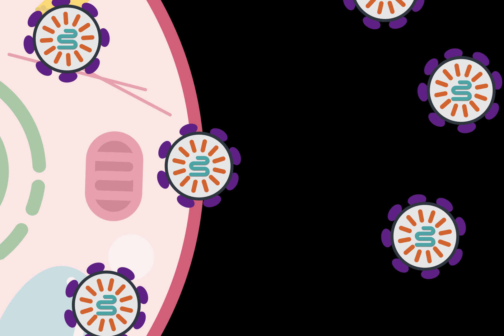
Each nanoparticle contained an applicable messenger RNA molecule that translates genetic information into functional proteins.

“We demonstrated two applications for lipid-like nanomaterials that effectively deliver their cargo, appropriately biodegrade, and are well-tolerated,” said Yizhou Dong, senior author of the study and associate professor of pharmaceutics and pharmacology at The Ohio State University.

“With this work, we have lowered potential side effects and toxicity and have broadened the therapeutic window. This gives us confidence to pursue studies in larger animal models and future clinical trials.”

This work builds upon a collection of lipid-like spherical compounds that Dong and colleagues had previously developed to deliver messenger RNA. This line of particles was designed to target disorders involving genes that are expressed in the liver.

The team experimented with various structural changes to those particles, effectively adding “tails” of different types of molecules to them, before landing on the structure that made the materials the most stable. The tiny compounds have a big job to do: embarking on a journey through the bloodstream, carrying molecules to the target location, releasing the ideal concentration of messenger RNA cargo at precisely the right time, and safely degrading.

The tests in mice suggested these particles could do just that.

The researchers injected nanoparticles containing messenger RNA holding the instructions to produce a protein called human factor VIII into the bloodstream of normal mice and mouse models for hemophilia. A deficiency of this protein, which enables blood to clot, causes the bleeding disorder. Within 12 hours, the deficient mice produced enough human factor VIII to reach 90 percent of normal activity. A check of the organs of both protein-deficient mice and normal mice showed that the treatment caused no organ damage.

“It can be helpful to think of this as a protein-replacement therapy,” Dong said.

In the second experiment, nanomaterials were loaded with two types of instructions: messenger RNA carrying the genetic code for a DNA base editor, and a guide RNA to make sure the edits occurred in a specific gene in the liver called PCSK9. Dozens of mutations that increase this gene’s activity are known to cause high cholesterol by reducing clearance of cholesterol from the bloodstream.

Analyses showed that the treatment resulted in the intended mutation of about 60 percent of the target base pairs in the PCSK9 gene, and determined that only a low dose was needed to produce high editing effect.

Dong credited academic and industry partners for helping advance this work. Co-corresponding authors include Denise Sabatino of Children’s Hospital of Philadelphia and Delai Chen from Boston-based Beam Therapeutics, who provided expertise in hemophilia and DNA base editing, respectively.

Dong and first author Xinfu Zhang are inventors on patent applications filed by Ohio State related to the lipid-like nanoparticles. This technology has been licensed for further clinical development.

This work was supported by the National Institute of General Medical Sciences, the National Heart, Lung and Blood Institute, and a startup fund from Ohio State’s College of Pharmacy.

Additional co-authors are Giang N. Nguyen of Children’s Hospital of Philadelphia; Weiyu Zhao, Chengxiang Zhang, Chunxi Zeng, Jingyue Yan, Shi Du, Xucheng Hou, Wenqing Li, Justin Jiang, Binbin Deng and David McComb of Ohio State; and Robert Dorkin, Aalok Shah, Luis Barrera, Francine Gregoire and Manmohan Singh of Beam Therapeutics.

Reference
Zhang X et al. Functionalized lipid-like nanoparticles for in vivo mRNA delivery and base editing. (2020) Sci Adv 6:eabc2315. doi: 10.1126/sciadv.abc2315. PMID: 32937374.