Glioblastoma multiforme is the most common type of malignant brain tumor. These tumors actively divide and send invasive cells throughout the brain. This migration complicates patient treatment, because simply removing the tumor does not clear the brain of migrating cells, which can initiate new tumors elsewhere. This problem has nearly halted progress in prognosis or treatment of glioblastoma over the past 20 years, and has afflicted many of our most important political figures, including Ted Kennedy and John McCain. This astonishing lack of improvement in treatment has motivated Dr. Xandra Breakefield and her team to explore an alternative pathway for treatment. Dr. Breakefield discussed her work in a recent Wednesday afternoon lecture at NIH.
To start their investigation, Dr. Breakefield gathered a “fresh” sample of glioblastoma multiforme that came straight out of the operating room. The idea of studying a fresh sample was novel; previously, samples were a few hours old or from mouse models. Examining fresh samples in a living condition revealed that glioblastomas are extremely physically active, constantly extending protrusions from the surface of the tumor cells. These protrusions turn into extracellular vesicles (EVs) of many sizes, released at the rate of about 10,000 vesicles per cell per day. When profiled, these EVs were found to contain RNAs, enzymes, transcription factors, other proteins, and many other cellular components. The team hypothesized that the EVs released by the tumor must somehow promote tumor progression.
Using a fluorescent tag that labels both cell and vesicle membranes, they tracked the tumor EVs in the living brain of mice and found that many of them are taken up by surrounding healthy myeloid cells — microglia and macrophages. Macrophages are the primary form of defense in the central nervous system. Normally functioning microglia are sentinels, warriors, and nurturers. As sentinels, they actively scan the brain for damage, then rush in to nurture injured areas to repair the damage. In their warrior role they kill and eat invading cells. Ironically, a higher density of microglia and macrophages in a tumor results in a worse prognosis. They are attracted to but then subjugated by the glioblastoma, and are coerced into an abnormal function of supporting tumor growth. Understanding how glioblastoma sabotages these immune cells and co-opts them to support tumor growth is key to ultimately finding a cure for the disease.
Glioblastomas and their EVs typically have very high levels of miR-21, a microRNA affiliated with various cancer pathways. Dr. Breakefield and her team found that microglia grown near a glioblastoma and exposed to its EVs had higher levels of miR-21, higher proliferation rates, and lower expression of proteins from pathways involved in sensing and attacking invaders. They hypothesized that miR-21 transferred from the tumor to surrounding benign microglia by EVs plays a key role in recruiting and transforming them. Then they asked whether microglia that have taken up many tumor vesicles have a different phenotype than microglia that haven’t taken up as many. To test this question, the Breakefield lab implanted fluorescently (GFP) labelled gliomas into the brains of mice and developed a flow cytometry method to sort cells extracted from that environment based on their level of GFP. The more tumor vesicles the surrounding normal cells have taken up, the more GFP they should have. The researchers used single-cell RNA sequencing to compare the expression profile of mRNAs in microglia and macrophages with high vs. low levels of GFP, indicating uptake of tumor vesicles. They found significant differences in mRNA expression in brain microglia but not in macrophages.
The genetic pathways responsible for microglia’s sentinel role have been called the “microglial sensome” (Hickman et al., 2013). The mRNA for most sensome genes were down-regulated in microglia that had taken up more tumor EVs – thus, the tumor EVs “blinded” the microglia. Pathways for immune suppression were up-regulated – showing that uptake of the tumor EVs compromised the microglias’ warrior function. Finally, pathways for tissue repair were up-regulated – indicating that the tumor EVs co-opted the microglias’ nurturing function to support the tumor instead of normal brain cells.
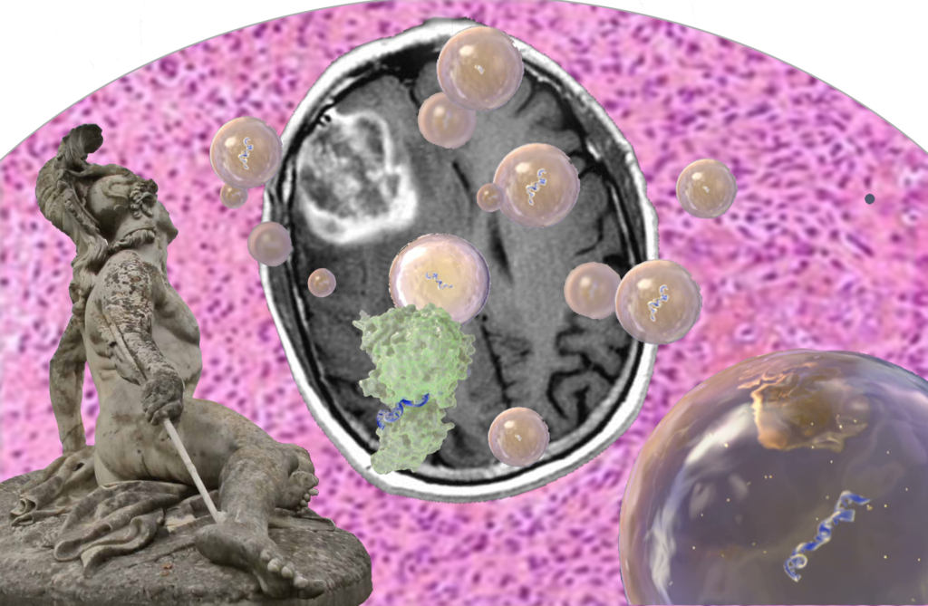
EVs are a promising biomarker for earlier diagnosis of disease. They are released by all cells and found in all biofluids. They can contain a wide variety of different information, including cell specific proteins and RNAs.
Dr. Breakefield was interested in finding biomarkers for glioblastoma that could be found in the bloodstream or other accessible biofluids. She targeted EGFRvIII, a deletion in the EGFR gene quite common in glioblastomas, and IDH1/2, a single point mutation that results in lower grade tumors with better prognosis. Her team is examining cerebrospinal fluid (CSF) and serum from patients to identify the presence or absence of EGFRvIII with promising success. Examining exRNA and free circulating DNA in plasma, her team was able to differentiate IDH1/2 mutant carriers from healthy volunteers. That work is being developed into a clinical tool for earlier diagnosis of low-grade gliomas which have a better prognosis and different treatment options than non-IDH1/2 gliomas.
Therapy EVs are proven, effective therapies for a variety of diseases. They can be obtained from many cell types and protect their fragile molecular cargo when administered into the body. They can also be efficiently taken up by specific target cells in vivo. EVs have been used therapeutically in immune modulation, tissue repair, and antigen presentation for vaccination.
After working on glioblastomas for an extended period, Dr. Breakefield decided to study schwannomas, a related but benign class of tumor. There are three types of hereditary diseases that stem from schwannomas: neurofibromatosis 1 (NF1), neurofibromatosis 2 (NF2), and schwannomatosis. These diseases cause motor dysfunction, pain, and potential hearing loss.
In NF2, tumors form along nerve fibers, including those at the base of the spine. The current treatment is surgical removal of the tumor, but that can cause irreparable nerve damage. Because these tumors are not malignant, reducing their size could be an effective alternative treatment.
Dr. Breakefield and colleagues made a model system using human Schwann cells from an NF2 patient. They implanted those cells in the sciatic nerve of mice, forming tumors. They then created an adenovirus-associated virus (AAV) vector containing the pro-inflammatory enzyme caspase 1 attached to a promoter (P0) exclusively active in Schwann cells. They then injected this vector into the tumors to try to shrink them.
In this model system, the tumor grew when the AAV-GFP was injected. It regressed when AAV-P0-ICE was injected. P0-ICE creates a “bystander” effect, in which cells surrounding the injected cell are also killed. Surprisingly, Schwann cells appeared completely normal after treatment with P0-ICE, while the tumor (schwannoma) shrunk after injection of the tumor with AAV-P0-ICE. Although the EVs’ role in this finding is not known, Dr. Breakefield theorizes that caspase-1 is incorporated into the EVs and transferred to surrounding schwannoma cells.
There is still much to be discovered about EVs’ roles in tumors, both benign and malignant. We have not yet found glioblastoma’s Achilles’ heel, but further research may yet do so. Dr. Breakefield looks forward to continuing her work and potentially creating better treatment options for those afflicted by these devastating tumors. The previously unrecognized role of the tumor-derived EVs in transferring genetic information and co-opting other cells in their microenviroment may be an important key.
Plasma Viral miRNAs Indicate a High Prevalence of Occult Viral Infections
Alterations in blood-based miRNA in veterans affected with combat-related PTSD

There are no comments.