Dr. Alissa Weaver, Vanderbilt University professor and Extracellular RNA Communication consortium (ERCC) member, will be inducted as an AAAS Fellow this Saturday, February 18, 2016. Dr. Weaver joins Dr. James Patton, also of Vanderbilt, and Dr. David Wong of UCLA as consortium members who are also current AAAS Fellows. This honor is bestowed upon her for her contributions to the field of cancer biology and studies of extracellular vesicles (EVs) in cell motility and cancer metastasis.
Dr. Weaver’s academic career began at Stanford University where she double majored in Biology and Political Science. Always aspiring to be a physician, she then attended medical school at the University of Virginia, Charlottesville. However, along the way, she realized that she missed the academics of a PhD. “When I was in medical school, I realized that I really missed thinking about scientific discovery and was not being taught to do research,” she explained. “I really wanted to have the formal training of getting a PhD so I applied for the program from medical school.” After completing her MD/PhD at UVA, she traveled to Washington University, Saint Louis for 5 years where she did a Laboratory Medicine residency and a postdoctoral fellowship in the Department of Cell Biology and Physiology with Dr. John Cooper.
Finally in 2003, she accepted a faculty position at Vanderbilt University where she now remains as a full time researcher. Her lab focuses on all aspects of extracellular vesicles. The interest originally stemmed from her investigations of cell invasion, migration and cancer metastasis. The lab’s focus shifted as they learned that many of the secreted molecules that facilitated invasion were transported by EVs.
Part of the invasive nature of cancer cells in metastasis involves structures called invadopodia, actin-based protrusions of the plasma membrane that facilitate degradation of the extracellular matrix. For cells to invade, they secrete matrix-degrading proteinases. Work in Weaver’s lab demonstrated that not only were these proteinases carried by EVs but that hot spots for their secretion actually aligned with invadopodia.
Specifically, Weaver’s lab established that invadopodia are important sites for the docking and secretion of exosomes. Exosomes are extracellular vesicles secreted from many different cell types. They originate from multivesicular bodies (MVB), which are mature endosomes that contain many smaller vesicles. Secretion of exosomes occurs when these MVBs fuse with the cell membrane, releasing the molecules contained inside. Though normal cells may use environmental cues to regulate exosome secretion, cancerous cells constitutively turn it on.
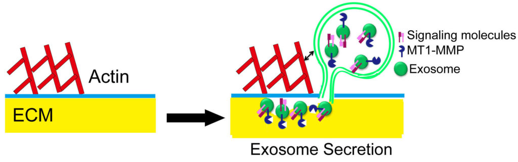
Exosome cargoes mediate invadopodia biogenesis, stability, and activity.
Source: Hoshino, et al. Cell Rep 2013
“One of the big questions we are working on is the cell biological aspects of these vesicles,” Weaver explained. “How they are made, how cargo gets sorted there, and what does that mean for their biological function after they are secreted? So that is where our work with the ERCC comes in.”
She hopes that working with the scientists of the consortium, they can understand how RNA and RNA binding proteins are trafficked into vesicles. Last year, in a paper published in Cell Reports, her group demonstrated one possible mechanism for the sorting of microRNAs into EVs. They demonstrated that Argonaute 2 (Ago2), part of the RISC machinery that binds to miRNAs, is transported in microvesicles and exosomes. Organization of Ago2 into exosomes is regulated by KRAS-MEK signaling. Dr. Weaver highlighted the study in a blog here mid-last year.
Despite these initial findings, Dr. Weaver admits it is difficult to determine how important extracellular RNA and miRNAs are in regulating cancer metastasis. “I honestly don’t think we know yet, and I think that the field is just now really trying to figure out what are the cargo components that are driving all of these phenotypes we have been trying to characterize so well.” She elaborated, “I think for both the protein and the RNA, the next big step for the field is trying to pin individual EV functions back to specific cargo molecules.”
Asked to reflect on her AAAS fellowship, Dr. Weaver turned the focus on her colleagues in the consortium. “I continue to be very impressed by the quality of investigators and the research being done by the ERCC. I mean really top people who are driving forward what I think is a tough problem.” She and fellow AAAS fellows Dr. Patton and Dr. Wong are, as Dr. Weaver pointed out, “just a small snapshot of fabulous investigators that are part of the consortium.”