News


Image: Kelsey Burke

This post originated as a press release from the University of Pennsylvania.

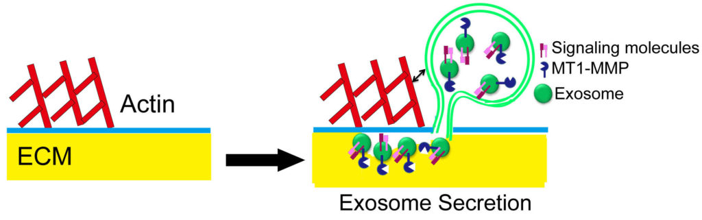
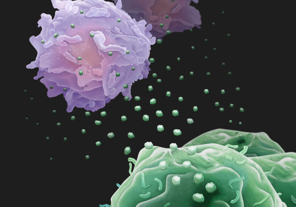
Cancerous tumors are more than a lump of cells growing out of control; they participate in active combat with the immune system for their own survival. Being able to evade the immune system is indeed a hallmark of cancer. Now, researchers from the University of Pennsylvania show that, to assist in the fight, cancer cells release biological “drones,” small vesicles called exosomes circulating in the blood and armed with the protein PD-L1, which causes T cells to tire before they have a chance to reach the tumor and do battle.

The work, published in the journal Nature (Chen et al., 2018), is a collaboration between Wei Guo of Penn’s School of Arts and Sciences and Xiaowei Xu of the Perelman School of Medicine. While primarily focused on metastatic melanoma, the team found that breast and lung cancer also release the PD-L1-carrying exosomes.

The research offers a paradigm-shifting picture of how cancers take a systemic approach to suppressing the immune system. In addition, it also points to a new way to predict which cancer patients will respond to certain checkpoint inhibitor drugs, which disrupt immune suppression to fight tumors, and a means of tracking the effectiveness of such therapies.

“Immunotherapies are life-saving for many patients with metastatic melanoma, but about 70 percent of these patients don’t respond,” says Guo, a professor of biology. “These treatments are costly and have toxic side effects, so it would be very helpful to know which patients are going to respond. Identifying a biomarker in the bloodstream could potentially help make early predictions about which patients will respond and, later on, could offer patients and their doctors a way to monitor how well their treatment is working.”

“Exosomes are tiny lipid-encapsulated vesicles with the diameter less than one-hundredth of a red blood cell. What we have found with these circulating exosomes is truly remarkable,” says Xu. “We collected blood samples from melanoma patients treated with anti-PD1 checkpoint inhibitor therapy. This type of liquid biopsy assay allows us to monitor tumor-related immune suppression with time.”

One of the most successful innovations in cancer therapy has been the use of checkpoint inhibitor drugs, which are designed to block attempts by cancer cells to suppress the immune system to allow tumors to thrive and spread. One of the primary targets for this class of drugs is PD-1, a protein on the surface of T cells. Tumor cells express PD-L1, which interacts with PD-1, effectively turning off that cell’s anti-cancer response. Blocking that interaction using checkpoint inhibitors reinvigorates T cells, allowing them to unleash their cancer-killing power on the tumor.

While it was known that cancer cells carried PD-L1 on their surface, Guo, Xu and colleagues found, in the new work, that exosomes from human melanoma cells also carried PD-L1 on their surface. Exosomal PD-L1 can directly bind to and inhibit T cell functions. Identifying the exosomal PD-L1 secreted by tumor cells provides a major update to the immune checkpoint mechanism, and offers novel insight into tumor immune evasion.

“Essentially exosomes secreted by melanoma cells are immunosuppressive.” Guo says. “We propose a model in which these exosomes act like drones to fight against T cells in circulation, even before the T cells get near to the tumor.”

Since a single tumor cell is able to secrete many copies of exosomes, the interaction between the PD-L1 exosomes and T cells provides a systemic and highly effective means to suppress anti-tumor immunity in the whole body. This may help explain why cancer patients have weakened immune systems.

Because exosomes circulate in the bloodstream, they present an accessible way of monitoring the cancer-T cell battle through a blood test, compared to a traditional, more-invasive tumor biopsy. After an acute phase of treatment, the researchers envision such a test as a way to monitor how well the drugs are keeping cancer cells in check.

By measuring pre-treatment levels of PD-L1, oncologists may be able to predict the extent of tumor burden in a patient and associate that with treatment outcome. In addition, a blood test could measure the effectiveness of a treatment. For example, levels of exosomal PD-L1 could indicate the level of T cell invigoration by immune checkpoint inhibitors.

“In the future, I think we will begin to think about cancers as a chronic disease, like diabetes,” says Guo. “And just as diabetes patients use glucometers to measure their sugar levels, it’s possible that monitoring PD-L1 and other biomarkers on the circulating exosomes could be a way for clinicians and cancer patients to keep tabs on treatment. It’s another step toward precision and personalized medicine.”


Reference

Chen G. et al. Exosomal PD-L1 contributes to immunosuppression and is associated with anti-PD-1 response. Nature (2018) 560: 382–386. doi: 10.1038/s41586-018-0392-8 PMID: 30089911


Guo and Xu coauthored the work with Penn’s Gang Chen, Alexander C. Huang, Wei Zhang, Min Wu, Jiegang Yang, Beike Wang, Honghong Sun, Wenqun Zhong, Bin Wu, Xiaoming Liu, Lei Guan, Tin Li, Shujing Liu, Ruifeng Yang, Youtao Lu, Liyun Dong, Suzanne McGettigan, Ravi Radhakrishnan, Junhyong Kim, Youhai H. Chen, Giorgos C. Karakousis, Tara C. Gangadhar, Lynn M. Schuchter, and E. John Wherry, as well as collaborators from Wuhan University, The Wistar Institute, Xi’an Jiaotong University, the University of Texas MD Anderson Cancer Center, and the Mayo Clinic.

The research was supported by the National Institutes of Health (GM111128, GM085146, AI105343, AI108545, AI082630, and AI117950), Parker Institute for Cancer Immunotherapy, American Heart Association, Tara Miller Melanoma Foundation, University of Pennsylvania, Wistar Institute, Dr. Miriam and Sheldon G. Adelson Medical Research Foundation, CAST Foundation, and NSFC Foundation.

Wei Guo is a professor of biology in the School of Arts and Sciences, and Xiaowei Xu is a professor of pathology and laboratory medicine and of dermatology in the Perelman School of Medicine at the University of Pennsylvania.

Wei Guo, Xiaowei Xu, and Gang Chen are listed as inventors on a patient owned by the University of Pennsylvania related to this work. Guo and Xu serve on the Scientific Advisory Board and have equities in Exo Bio, a company that has licensed the patent from the University of Pennsylvania.

This post originated as a press release from Linköping University.

The waste-management system of the cell appears to play an important role in the spread of Alzheimer’s disease in the brain. A new study, published in the prestigious scientific journal Acta Neuropathologica, has focused on small membrane-covered droplets known as exosomes. It was long believed that the main task of exosomes was to help the cell to get rid of waste products. In simple terms, they were thought of as the cell’s rubbish bags. However, our understanding of exosomes has increased, and we now know that cells throughout the body use exosomes to transmit information. It’s now known that the exosomes can contain both proteins and genetic material, which other cells can absorb.

The Linköping researchers have shown in the new study that exosomes can also transport toxic aggregates of the protein amyloid beta, and in this way spread the disease to new neurons. Aggregated amyloid beta is one of the main findings in the brains of patients with Alzheimer’s disease, the other being aggregates of the protein tau. As time passes, they form ever-increasing deposits in the brain, which coincides with the death of nerve cells. The cognitive functions of a person with Alzheimer’s disease gradually deteriorate as new parts of the brain are affected.

“The spread of the disease follows the way in which parts of the brain are anatomically connected. It seems reasonable to assume that the disease is spread through the connections in the brain, and there has long been speculation about how this spread takes place at the cellular level,” says Martin Hallbeck, associate professor in the Department of Clinical and Experimental Medicine at Linköping University and senior consultant of clinical pathology at Linköping University Hospital.

Cells became diseased
In a collaboration with researchers at Uppsala University, he and his co-workers have investigated exosomes in brain tissue from deceased persons. The research team at Linköping University found more amyloid beta in exosomes from brains affected by Alzheimer’s disease than in healthy controls. Furthermore, the researchers purified exosomes from the brains from people with Alzheimer’s disease, and investigated whether they could be absorbed by cells cultured in the laboratory.

“Interestingly, exosomes from patients were absorbed by cultured neurons, and subsequently passed on to new cells. The cells that absorbed exosomes that contained amyloid beta became diseased,” says Dr. Hallbeck.

The researchers treated the cultured neurons with various substances that prevent exosomes from being formed, released, or absorbed by other cells. They were able to reduce the spread of the aggregated amyloid beta between cells by disrupting the mechanism in these ways. The methods used in these laboratory experiments are not yet suitable for treating patients, but the discovery is important in principle.

“Our study demonstrates that it is possible to influence this pathway, and possibly develop drugs that could prevent the spreading. The findings also open up the possibility of diagnosing Alzheimer’s disease in new ways, by measuring the exosomes,” says Martin Hallbeck.

The research has received financial support from donors that include the Swedish Research Council, the Swedish Alzheimer’s Foundation, and the Swedish Brain Foundation.

Sinha MS, Ansell-Schultz A, Civitelli L, Hildesjö C, Larsson M, Lannfelt L, Ingelsson M & Hallbeck M. Alzheimer disease pathology propagation by exosomes containing toxic amyloid-beta oligomers. Acta Neuropathologica AOP 13 June 2018. doi: 10.1007/s00401-018-1868-1

Translation by George Farrants.

Despite being one of the earliest known classes of non-coding RNA molecules, transfer RNAs (tRNAs) are still notoriously difficult to study. The challenge is largely due to this molecule’s secondary structure, chemical modifications to its constituent nucleotides (see figure), and the multiplicity of tRNA genes. As the number of non-coding RNA datasets proliferates, it is becoming increasingly important for tRNA genes to be accurately annotated. In a recent study, Thomas Tuschl from Rockefeller University and colleagues tackled this problem by developing a new protocol for sequencing tRNAs. The new method enabled them to assemble an atlas of human tRNAs for other researchers to use in analyzing their non-coding RNA data.

Hydro-tRNA Sequencing
Transfer RNAs have thermodynamically stable secondary and tertiary structures, and their constituent nucleotides are highly modified by RNA editing. Both of these characteristics are problematic for traditional RNA sequencing methods. The key to the Tuschl lab’s protocol, called hydro-tRNA sequencing (hydro-tRNAseq), is a partial alkaline hydrolysis step that breaks the 60-100 nucleotide-long tRNA into smaller fragments with fewer RNA modifications. These fragments, 19-35 nucleotides in size, have weaker secondary structure and fewer RNA modifications per fragment than the parent tRNA.

Applying the method to short RNA extracted from human embryonic kidney (HEK293) cells resulted in an increase in the fraction of reads mapped to tRNA between 2% and 40%, depending on the depth of sequencing. The short fragment length also improved read accuracy per base compared to standard tRNA sequencing.

To develop a thorough and representative reference set of human tRNAs, the HEK293 dataset was subjected to iterative cycles of mapping to existing reference tRNAs followed by manual curation. In each round, all transcripts with an error distance (number of mismatches, insertions, and deletions) of 1-2 from a given tRNA were kept as candidate reference sequences if they could be attributed to a tRNA isoacceptor (i.e. a different tRNA that binds to the same amino acid). If not, assuming that other mismatches were caused by misidentifying a modified base, transcripts with more than 10% mismatches compared to reference were expanded into a set of all possible combinations of RNA modifications and included in the reference pool (see figure). This mapping and selection process was repeated until there were no longer any modified positions left with a mismatch frequency over 10% compared to reference.

Candidate pre-tRNA genes were obtained by mapping the final tRNA reference sequences back to the genome. Altogether, this analysis was able to account for 93% of the 114 million reads in the deepest library of HEK293 cells’ tRNAs.

tRNA Modification Sites

tRNA Modification Sites
The team identified sites of modification from the high frequency of mismatches during mapping caused by read errors there during reverse transcription. Here the reference nucleotide is at ring center, known modification outside the ring, and frequency of each nucleotide read at that site inside the ring.
Source: Cell Reports

The Added Power of SSB PAR-CLIP
Though hydro-tRNAseq greatly improved the reference dataset of human tRNAs, there was still a risk that it alone would miss pre-tRNAs expressed at low levels or processed quickly into mature tRNA. Previous efforts to assay that ephemeral population employed ChIP-seq of POLR3, the polymerase that transcribes all tRNA genes, but doing so assumed that polymerase binding always led to expression and complete processing. The Tuschl lab focused instead on SSB, a protein that binds to the 3′ end of pre-tRNAs, immunoprecipitating tRNAs crosslinked to SSB using a method called PAR-CLIP. As predicted, almost half of the reads from their SSB PAR-CLIP experiments mapped to pre-tRNAs. Combining SSB PAR-CLIP with hydro-tRNAseq allowed the team to better identify mature and pre-tRNAs with improved, accurate, nucleotide-level resolution.

This study supplies the community with several new and useful resources. Hydro-tRNAseq provides a new method to overcome many of the struggles of tRNA sequencing analyses. Combining this method with SSB PAR-CLIP enabled the construction of a comprehensive atlas of pre-tRNAs and mature tRNAs in humans. This methodology can now be applied to study the tRNA complement in other species to further dissect tRNA biology.

Reference
Tasos Gogakos T, Brown M, Garzia A, Meyer C, Hafner M, & Tuschl T. Characterizing Expression and Processing of Precursor and Mature Human tRNAs by Hydro-tRNAseq and PAR-CLIP. Cell Reports (2017) 20: 1463-1475. doi: 10.1016/j.celrep.2017.07.029

(This blog first appeared as a press release from Ohio State University.)

Principal investigator Peixuan Guo, PhD, Sylvan G. Frank Endowed Chair professor of the OSU College of Pharmacy and a member of the OSUCCC – James Translational Therapeutics Program.

  • Therapies based on RNA, such as small interfering RNA, hold great promise for cancer treatment but delivering these agents to their targets in cancer cells has been a problem.
  • A new study shows that attaching antibody-like RNA nanoparticles to microvesicles can deliver effective RNA therapeutics specifically to cancer cells.
  • The researchers are now working to adapt the technology for use in the clinic.

Columbus, Ohio – A new study shows that attaching antibody-like RNA nanoparticles to microvesicles can deliver effective RNA therapeutics such as small interfering RNA (siRNA) specifically to cancer cells. Researchers used RNA nanotechnology to apply the RNA nanoparticles and control their orientation to produce microscopic, therapy-loaded extracellular vesicles that successfully targeted three types of cancer in animal models.

The findings, reported in the journal Nature Nanotechnology, could lead to a new generation of anticancer drugs that use siRNA, microRNA and other RNA-interference technologies.

The study was led by researchers at Ohio State’s College of Pharmacy; the Ohio State University Comprehensive Cancer Center – James Cancer Hospital and Solove Research Institute (OSUCCC – James).

“Therapies that use siRNA and RNA interference technologies are poised to transform cancer therapy,” says the principal investigator Peixuan Guo, PhD, Sylvan G. Frank Endowed Chair professor of the College of Pharmacy and a member of the OSUCCC – James Translational Therapeutics Program. “But clinical trials evaluating these agents have failed one after another due to the inability to deliver the agents directly to cancer cells in the human body.”

Guo noted that even when agents did reach and enter cancer cells, they were trapped in internal vesicles called endosomes and rendered ineffective.

“Our findings solve two major problems that impede these promising anticancer treatments: targeted delivery of the vesicles to tumor cells and freeing the therapeutic from the endosome traps after it is taken up by cancer cells. In this study, cancers stopped growing after systemic injection of these particles into animal models with tumors derived from human patients.” Guo says. “We’re working now to translate this technology into clinical applications.”

Guo and his colleagues produced extracellular microvesicles (exosomes) that display antibody-like RNA molecules called aptamers that bind with a surface marker that is overexpressed by each of three tumor types:

  • To inhibit prostate cancer, vesicles were designed to bind to prostate-specific membrane antigen (PSMA);
  • To inhibit breast cancer, vesicles were designed to bind to epidermal growth factor receptor (EGFR);
  • To inhibit a colorectal cancer graft of human origin, vesicles were designed to bind to folate receptors.

All vesicles were loaded with a small interfering RNA for down-regulating the survivin gene as a test therapy. The survivin gene inhibits apoptosis and is overexpressed in many cancer types.

Key findings include:

  • Vesicles targeting the prostate-specific membrane antigen completely inhibited prostate-cancer growth in an animal model with no observed toxicity.
  • Vesicles targeting EGFR inhibited breast cancer growth in an animal model.
  • Vesicles targeting folate receptors significantly suppressed tumor growth of human patient-derived colorectal cancer in an animal model.

“Overall, our study suggests that RNA nanotechnology can be used to program natural extracellular vesicles for delivery of interfering RNAs specifically to cancer cells,” Guo says.

Funding from the National Institutes of Health/National Cancer Institute (grants TR000875 and CA207946, CA186100, CA197706, CA177558 and CA195573) supported this research.

Other researchers involved in this study were Fengmei Pi, Daniel W. Binzel, Zhefeng Li, Hui Li, Farzin Haque, Shaoying Wang and Carlo M. Croce, The Ohio State University Wexner Medical Center; Meiyan Sun and Bin Guo, University of Houston; Piotr Rychahou and B. Mark Evers, University of Kentucky; and Tae Jin Lee, now at University of Texas.

About the OSUCCC – James
The Ohio State University Comprehensive Cancer Center – Arthur G. James Cancer Hospital and Richard J. Solove Research Institute strives to create a cancer-free world by integrating scientific research with excellence in education and patient-centered care, a strategy that leads to better methods of prevention, detection and treatment. Ohio State is one of only 49 National Cancer Institute (NCI)-designated Comprehensive Cancer Centers and one of only a few centers funded by the NCI to conduct both phase I and phase II clinical trials on novel anticancer drugs sponsored by the NCI. As the cancer program’s 308-bed adult patient-care component, The James is one of the top cancer hospitals in the nation as ranked by U.S. News & World Report and has achieved Magnet designation, the highest honor an organization can receive for quality patient care and professional nursing practice. At 21 floors with more than 1.1 million square feet, The James is a transformational facility that fosters collaboration and integration of cancer research and clinical cancer care.


Scientists from the ERCC have joined forces to create a CSF Consortium to pool resources and establish standard practices in the study of cerebrospinal fluid (CSF).

One of the goals of the ERCC is not only to understand the fundamental biology of extracellular RNA (exRNA), but to develop exRNA-based biomarkers of disease. When such biomarkers have been found, studied, and cleared for clinical use, liquid biopsy of blood and other biofluids can enable earlier disease detection and less invasive tracking of disease progression. For neurological disorders, drawing CSF from the spinal cord has clear benefits over a more invasive brain biopsy. Progress in our technical understanding of how to accurately assess biomarkers in CSF will increase our basic understanding and promote clinical advancements in the diagnosis and treatment of neurological disease. Unfortunately, there are many inconsistencies between the processing of CSF in current studies. Data replication is often difficult, in large part due to variability across laboratories and institutions in protocols for sample isolation, purification, and analysis. Thus, the CSF Consortium, spearheaded by Dr. Fred Hochberg (https://fredhhochbergmd.com), was designed to be a resource for researchers to help minimize these discrepancies.

The CSF consortium plan calls for CSF researchers and clinicians to work together to improve standard practices. A major focus is transparency through open sharing of their work. Researchers are encouraged to establish collaborations, share in-depth details of experimental designs and reagents (including batch/lot numbers), and release any details of in-house protocol modifications. Working with the same biosamples shared through the Virtual Biorepository (VBR) enables multiple labs to compare and synchronize their protocols with one source of variability removed. The expectation is that sharing of detailed information will enable future researchers to avoid common pitfalls and plan their own experiments appropriately. Ultimately, the goal is to have open-access information available from each stage of every project: from biofluid, RNA, and extracellular vesicle (EV) collection, isolation, and storage to downstream analyses such as RT-qPCR and RNA sequencing.

If you are a CSF researcher, please contact us so that we can work with you as well!

Highlights of recent CSF Consortium efforts
Saugstad et al. (2017) recently demonstrated the strength of the CSF consortium. In a collaboration between three institutions (UC San Diego, Oregon Health & Science University, and the Translational Genomics Research Institute), researchers worked together to characterize the EV and RNA composition of identical pools of CSF at each institute from patients with five different neurological disorders. This work in parallel allowed the groups to identify potential sources of variability in protocols including sample preparation, RNA isolation, and quantification of RNA via RNA sequencing and RT-qPCR. The study identified changes in EVs and RNA in the disease CSF samples and detected an enrichment of microRNAs and mRNAs related to disease in both EV and total RNA. The paper highlights the importance of stringent standard operating procedures, including the use of common standard sample collection and data analysis protocols across institutions.

In other work, Figueroa et al., 2017 performed a multi-institutional study of RNA extracted from CSF-derived EVs of patients with glioblastoma (GBM), a very aggressive form of brain cancer. (See this related blog on glioblastoma.) A key diagnostic biomarker in classical GBM is the functional status of the Epidermal Growth Factor Receptor (EGFR). This cell-surface receptor is the starting point of a series of signaling pathways related to cell growth. When its expression surges or it folds incorrectly, the result is cells with hyper-active signaling that never stop growing. This study involved the development of a liquid biopsy that scans RNA extracted from CSF EVs for tumor-associated amplifications and mutations in EGFR. The test has very high specificity and fair sensitivity: it almost never incorrectly flags a healthy patient as having GBM and correctly identifies almost two thirds of GBM sufferers. The clinical standard for diagnosis of GBM is magnetic resonance imaging (MRI), which correctly classifies most brain tumors, but in too many cases incorrectly suggests that healthy brain tissue might be cancerous. The complementarity of highly sensitive MRI and highly specific RNA liquid biopsy argues that updating the standard of care to include collection of CSF and brain images at the same time would better separate healthy from diseased brain tissue.

The CSF Consortium is casting its nets wider in its fight against glioblastoma, looking at molecules beyond EGFR in the attempt to develop an RNA-based diagnosis tool for GBM. Akers et al., 2017 developed a diagnostic panel of 9 miRNA biomarkers by analyzing the EV RNA from 135 CSF samples in 3 cohorts, followed by validation of the miRNA panel in 60 CSF samples from 2 cohorts. The researchers found that even with that fairly large sample size, the miRNA profiles in lumbar and cisternal CSF — fluid collected from the spine or the base of the neck, respectively — are significantly different, which is problematic, since cisternal CSF is much more difficult to collect. On the other hand, they also found that RNAs extracted from raw CSF had a similar profile and diagnostic power as RNAs extracted from vesicles after an initial EV purification step, which might simplify the translation of this biomarker research into the clinic.

References
Akers, J.C. et al. A cerebrospinal fluid microRNA signature as biomarker for glioblastoma. Oncotarget (2017) 8: 68769-68779.
Figueroa, J.M et al. Detection of wtEGFR amplification and EGFRvIII mutation in CSF-derived extracellular vesicles of glioblastoma patients. Neuro. Oncol. (2017) Advance online publication. doi: 10.1093/neuonc/nox085
Saugstad, J.A. et al. Analysis of extracellular RNA in cerebrospinal fluid. J. Extracellular Vesicles (2017) 6: 1317577.

Dr. Alissa Weaver, Vanderbilt University professor and Extracellular RNA Communication consortium (ERCC) member, will be inducted as an AAAS Fellow this Saturday, February 18, 2016. Dr. Weaver joins Dr. James Patton, also of Vanderbilt, and Dr. David Wong of UCLA as consortium members who are also current AAAS Fellows. This honor is bestowed upon her for her contributions to the field of cancer biology and studies of extracellular vesicles (EVs) in cell motility and cancer metastasis.

Alissa Weaver

Dr. Weaver’s academic career began at Stanford University where she double majored in Biology and Political Science. Always aspiring to be a physician, she then attended medical school at the University of Virginia, Charlottesville. However, along the way, she realized that she missed the academics of a PhD. “When I was in medical school, I realized that I really missed thinking about scientific discovery and was not being taught to do research,” she explained. “I really wanted to have the formal training of getting a PhD so I applied for the program from medical school.” After completing her MD/PhD at UVA, she traveled to Washington University, Saint Louis for 5 years where she did a Laboratory Medicine residency and a postdoctoral fellowship in the Department of Cell Biology and Physiology with Dr. John Cooper.

Finally in 2003, she accepted a faculty position at Vanderbilt University where she now remains as a full time researcher. Her lab focuses on all aspects of extracellular vesicles. The interest originally stemmed from her investigations of cell invasion, migration and cancer metastasis. The lab’s focus shifted as they learned that many of the secreted molecules that facilitated invasion were transported by EVs.

Part of the invasive nature of cancer cells in metastasis involves structures called invadopodia, actin-based protrusions of the plasma membrane that facilitate degradation of the extracellular matrix. For cells to invade, they secrete matrix-degrading proteinases. Work in Weaver’s lab demonstrated that not only were these proteinases carried by EVs but that hot spots for their secretion actually aligned with invadopodia.

Specifically, Weaver’s lab established that invadopodia are important sites for the docking and secretion of exosomes. Exosomes are extracellular vesicles secreted from many different cell types. They originate from multivesicular bodies (MVB), which are mature endosomes that contain many smaller vesicles. Secretion of exosomes occurs when these MVBs fuse with the cell membrane, releasing the molecules contained inside. Though normal cells may use environmental cues to regulate exosome secretion, cancerous cells constitutively turn it on.

Exosome cargoes mediate invadopodia biogenesis, stability, and activity

Exosome cargoes mediate invadopodia biogenesis, stability, and activity.
Source: Hoshino, et al. Cell Rep 2013

“One of the big questions we are working on is the cell biological aspects of these vesicles,” Weaver explained. “How they are made, how cargo gets sorted there, and what does that mean for their biological function after they are secreted? So that is where our work with the ERCC comes in.”

She hopes that working with the scientists of the consortium, they can understand how RNA and RNA binding proteins are trafficked into vesicles. Last year, in a paper published in Cell Reports, her group demonstrated one possible mechanism for the sorting of microRNAs into EVs. They demonstrated that Argonaute 2 (Ago2), part of the RISC machinery that binds to miRNAs, is transported in microvesicles and exosomes. Organization of Ago2 into exosomes is regulated by KRAS-MEK signaling. Dr. Weaver highlighted the study in a blog here mid-last year.

Despite these initial findings, Dr. Weaver admits it is difficult to determine how important extracellular RNA and miRNAs are in regulating cancer metastasis. “I honestly don’t think we know yet, and I think that the field is just now really trying to figure out what are the cargo components that are driving all of these phenotypes we have been trying to characterize so well.” She elaborated, “I think for both the protein and the RNA, the next big step for the field is trying to pin individual EV functions back to specific cargo molecules.”

Asked to reflect on her AAAS fellowship, Dr. Weaver turned the focus on her colleagues in the consortium. “I continue to be very impressed by the quality of investigators and the research being done by the ERCC. I mean really top people who are driving forward what I think is a tough problem.” She and fellow AAAS fellows Dr. Patton and Dr. Wong are, as Dr. Weaver pointed out, “just a small snapshot of fabulous investigators that are part of the consortium.”

Immunology 2016
Immunology 2016

Extracellular RNA was a hot topic of discussion at Immunology 2016, the annual meeting of the American Association of Immunologists (AAI), held at the Washington State Convention Center in Seattle, Washington May 13-17th, 2016. The National Cancer Institute (NCI) sponsored a symposium on “Extracellular RNA in the Immune System”, co-chaired by Dr. Kevin Howcroft (Division of Cancer Biology, Cancer Immunology, Hematology, and Etiology Branch, NCI) and K. Mark Ansel (University of California San Francisco – your faithful blogger). Four invited speakers presented and participated in lively discussion with an audience of gathered experts and curious newcomers to the field of extracellular RNA.

Dr. Gyongyi Szabo (University of Massachusetts) opened the symposium with a presentation of her laboratory’s work on extracellular vesicles and miRNAs in innate immune cell communication in the liver. Alcohol exposure induces liver inflammation, marked by release of pro-inflammatory cytokines and activation of myeloid cells, including Kupffer cells, the resident macrophages of the liver. In a mouse model, alcohol consumption increased expression of miR-155 in both macrophages and hepatocytes via TLR4 and NFκB-driven transcription. Inhibition or genetic deletion of miR-155 in this model blunted macrophage activation and cytokine production. Exosomes loaded with miR-155 mimetics could be delivered to hepatocytes and other liver cells to correct some of the defects observed in miR-155-deficient animals. Remarkably, endogenous miR-155 and miR-122 were elevated in serum collected after controlled “binge-drinking” in human study subjects, and these exosomes also conveyed information to cultured monocytes, altering their production of TNF and IL-1. Together these data suggest that extracellular communication between hepatocytes and innate immune cells via exosomal miRNAs regulates inflammation in response to alcohol consumption.

The theme of regulation of inflammatory responses by miRNA-containing exosomes was extended by Dr. Ryan O’Connell (University of Utah). His pioneering work on miR-155 and miR-146 demonstrated their opposing roles in inflammatory processes mediated by various cell types in several tissues and disease settings. Recent work in his laboratory showed that both of these miRNAs are released by bone-marrow-derived dendritic cells in a fashion dependent on Rab27 and neutral sphingomyelinase (N-SMase) activity, and that these miRNAs could be exchanged between cells separated by a filter that prevents cell-cell contact. Transferred miR-146a reduced recipient cells’ response to bacterial lipopolysaccharide, a classical innate immune stimulant in vitro and in vivo. In addition, transferred miR-155 was found to directly repress the 3’ UTR of target genes in recipient cells, supporting the possibility that functional miRNA transfer via exosomes could be used as a therapeutic modality for regulating inflammation. Getting these miRNAs to the right cell types in vivo remains an important challenge to bringing this technology to the clinic.

In addition to exosomes, high density lipoprotein (HDL) particles carry miRNAs and other extracellular RNAs in blood. Abnormal pro-inflammatory HDL is associated with systemic lupus erythematosus (SLE). Dani Michell (Vanderbilt University), a postdoctoral fellow in Kasey Vickers’ laboratory, discussed her work, conducted in collaboration with Amy Major’s laboratory, on miRNAs in HDL in SLE. HDL from subjects with SLE contained increased levels of miR-22-3p and miR-192-5p compared with HDL from healthy control subjects. Blocking miR-22 with locked nucleic acid inhibitors in vivo reduced spleen size and interferon production, and affected some clinical features in a mouse model of lupus. Experiments aimed at defining source and recipient cells in this system indicated that monocytes are much better than T lymphocytes at taking up HDL-associated miRNAs. It will be interesting to learn how HDL-associated miRNAs regain gene regulatory function in recipient cells.

The final presentation focused on lymphocytes as source cells for naturally occurring exRNAs in body fluids. Immuno-compromised mice with a mutation that specifically blocks lymphocyte development exhibit altered serum extracellular miRNA profiles. In support of the idea that lymphocytes themselves are an important source of ex-miRNAs, the most reduced exRNA species detected was miR-150, a miRNA highly expressed by lymphocytes. Activated T lymphocytes secrete vesicles that are enriched for tRNA fragments and miRNAs including miR-150. Rigorous purification revealed that these vesicles have characteristics of exosomes, including defined density, size, and protein markers including the tetraspanin CD9. Cellular fractionation also revealed tRNA fragment and miRNA enrichment in membrane fractions containing multivesicular bodies. Whether these extracellular lymphocyte-derived RNAs mediate cell-to-cell communication or not, signal-mediated reduction of cellular miRNAs certainly alters gene regulation in activated T lymphocytes. Thus, exRNA secretion may have important roles in regulating inflammatory processes in both source and recipient cells.

These topics will certainly remain on the mind of immunologists that attended the exRNA symposium — at least until Immunology 2017, to be held in Washington DC next May.

Non-coding RNAs (ncRNAs), for example microRNAs (miRNAs), are frequently dysregulated in cancer and other diseases, and have shown great potential as tissue-based markers for cancer classification and prognostication. ncRNAs are present in membrane-bound vesicles, such as exosomes, in extracellular human body fluids. Circulating miRNAs are also present in human plasma and serum and cofractionate with the Argonaute2 (Ago2) protein and high-density lipoprotein (HDL). Since miRNAs and other ncRNAs circulate in the bloodstream in highly stable forms, they may be used as blood-based biomarkers for cancer and other diseases. A knowledge base of non-invasive biomarkers is a fundamental tool for biomedical research in this field.

In 2012, miRandola was developed as the first database of circulating extracellular miRNAs (Russo et al., 2012). miRandola is a comprehensive, manually curated collection and classification of circulating extracellular miRNAs. We recently updated miRandola with 271 papers, 2695 entries, 673 miRNAs and 12 long non-coding RNAs. The future direction of the database is to be a resource for all potential non-invasive circulating nucleic acid biomarkers.

miRandola_schema

miRandola is the first online resource which gathers all the available data on circulating RNAs into one environment (see Figure). It represents a useful reference tool for anyone investigating the role of extracellular RNAs as biomarkers, as well as their physiological function and their involvement in pathologies.

The database is constantly updated as soon as new data is available, and the online submission system is a crucial feature which helps to ensure that the system is always up-to-date. We are working on a second version of the database to increase the amount of data and to improve usability. miRandola is available online at https://mirandola.iit.cnr.it/.

The Extracellular RNA Communication (ERC) Consortium Data Management and Resource Repository (DMRR) has released the latest version of the exRNA Atlas (BETA). This release contains preliminary data generated by the consortium and analyzed using the exceRpt small RNA-seq pipeline.

Key features of this release include:

  • Searches
    • Faceted search of exRNA profiles across biofluids, diseases or exRNA isolation method.
    • Drill-down subsetting of analyzed biosamples using interactive sunburst and linear tree diagrams.
    • Biosample partition grids with tabular views of biosamples collected and profiled for exRNAs from a biofluid/disease/experiment combination.
  •  

  • Summaries
    • Grid view of all studies submitted to the Data Coordination Center (DCC).
    • Barchart summaries of exRNA profiling datasets.
    • Tool usage summary grid displaying usage of exRNA profiling data analysis tools by ERC consortium members as well as other members of the scientific community.

Currently, the search and summary views in the Atlas can be accessed only by ERC Consortium members. If you are unable to login, please contact the Data Coordination Center for assistance.

A public version of the exRNA Atlas will be released next month.

Links

Watch a video tutorial highlighting all features in the current release of the exRNA Atlas.

Want to share your knowledge of a particular exRNA gene with the broader scientific community? Want to reach readers through both the “traditional” peer-reviewed literature as well as the sixth-most accessed website in the world? If so, continue reading about our three-way partnership between the journal GENE, the Gene Wiki project at Wikipedia, and the Extracellular RNA Communication Program (ERCP):

Gene_Wiki_logo

  1. What is it? The goal of the Gene Wiki is to create a comprehensive Wikipedia article for every human gene. To incentivize authors to improve Wikipedia content, GENE is now soliciting new gene-specific review articles under a new dual-publication model. ExRNA genes are especially desirable and authors are invited to create two separate versions of their review (one for the journal, and one in wikipedia). More on the partnership here: Gene Wiki Reviews: Marrying crowdsourcing with traditional peer review.
  2. How long should the review article be? The length of the review article is up to you! Since you are the expert on the exRNA gene you’re writing about, the length is based on whatever you think is necessary to describe the current state of the field.
  3. How long should the wikipedia article be? We are targeting a final length of approximately 1200 words (though longer and more detailed articles are certainly welcome)
  4. How are the two versions different? One version is targeted at professional scientists following typical academic and editorial standards. The second version is written for the Wikipedia audience and includes a slightly heavier emphasis on a general audience. Both versions will be peer-reviewed together, but for copyright reasons, these two versions must be separate works that have no substantial similarity. Some examples of review articles and wikipedia entries published under this model include:
  5. I am busy but intrigued, what is the time line? We generally suggest a 2-3 month deadline, but since this is an ongoing series in the journal, the time line is flexible and can be worked around your schedule. Don’t be discouraged from participating because you are busy now. Make the commitment to submit when your schedule permits.
  6. Do I have to go at this alone? Absolutely not! If you have colleagues who would make good co-authors for the review, feel free to solicit their assistance.
  7. Do I have to write the wiki article all at once? Nope. Our goal is to incentivize you, the expert, to make your knowledge about your exRNA gene accessible. If it’s easier for you to write the wiki article in pieces, go ahead and do so! As long as the wiki entry is complete by the time you submit your manuscript, we will be happy to accept your review article.
  8. The gene I work on doesn’t make much sense to write about alone, how should I contribute? Genes that work in concert can be tackled as a pair as with this example:
  9. Why should I do this? By publishing an exRNA gene-specific review article, you help your scientific colleagues stay abreast of the current literature on your favorite gene. By publishing under the dual publication model (ie- on wikipedia), you help make your favorite exRNA gene more accessible to everyone allowing more people to understand the importance of your field of research. Everyone wins!
  10. How do I get in on this? Check to see whether or not your favorite exRNA gene could use some serious contributions on wikipedia. If so, contact me. Include your exRNA gene of interest in the email, and your preferred deadline for the manuscript submission.

Looking forward to hearing from you!