This blog originated as a press release from Cedars-Sinai. Thanks to them for allowing us to repost it here.
A recent study sheds light on proteins in particles called extracellular vesicles, which are released by tumor cells into the bloodstream and promote the spread of cancer. The findings suggest how a blood test involving these vesicles might be used to diagnose cancer in the future, avoiding the need for invasive surgical biopsies.
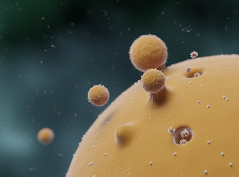 |
Extracellular vesicles Credit: NIH |
The research is a large-scale analysis of what are known as palmitoylated proteins inside extracellular vesicles, according to Dolores Di Vizio, MD, PhD, professor of Surgery, Biomedical Sciences and Pathology and Laboratory Medicine at Cedars-Sinai. Di Vizio is co-corresponding author of the study, published online June 10 in the Journal of Extracellular Vesicles.
Extracellular vesicles have gained significant attention in the last decade because they contain proteins and other biologically important molecules whose information can be transferred from cell to cell. They are known to help cancer metastasize to distant sites in the body, but exactly how this happens is not clear.
To learn more about this process, the research team looked into a process called palmitoylation, in which enzymes transfer lipid molecules onto proteins. Palmitoylation can affect where proteins are located within cells, their activities, and their contribution to cancer progression.
The investigators examined two types of extracellular vesicles, small and large, in samples of human prostate cancer cells. Using centrifuges, they separated the extracellular vesicles from the other cell materials and analyzed the levels of palmitoylation and the types of proteins present.
The team found extracellular vesicles derived from the cancer cells contained palmitoylated proteins that are associated with the spread of cancer. Further, when the team chemically suppressed the palmitoylation process, the level of some of these proteins went down in the extracellular vesicles.
“Our results suggest that protein palmitoylation may be involved in the selective packaging of proteins to different extracellular vesicle populations in the body,” Di Vizio said. “This finding raises the possibility that by examining these proteins in extracellular vesicles in the bloodstream, we may be able to detect and characterize cancer in a patient in the future without performing a surgical biopsy.”
 |
Dolores Di Vizio, MD, PhD Professor of Surgery, Biomedical Sciences and Pathology and Laboratory Medicine at Cedars-Sinai. |
Di Vizio said the next step in the research is to conduct a study in collaboration with her Cedars-Sinai colleagues and industry partners that will use advanced technologies, including mass spectrometry and flow cytometry, with the goal of identifying clinically significant prostate cancer at diagnosis.
In addition to Di Vizio, Wei Yang, PhD, associate professor of Surgery at Cedars-Sinai, and Andries Zijlstra, PhD, are co-corresponding authors for the study. Zijlstra completed the research while working at Vanderbilt University Medical Center in Nashville. Javier Mariscal, PhD, a postdoctoral scientist in Di Vizio’s laboratory, is the study’s first author.
Reference
Mariscal J et al. Comprehensive palmitoyl-proteomic analysis identifies distinct protein signatures for large and small cancer-derived extracellular vesicles. (2020) J Extracell Vesicles 9:1764192. doi: 10.1080/20013078.2020.1764192. PMID: 32944167.
Funding: Research reported in this publication was supported by the National Institutes of Health under award number R01CA218526 and by the U.S. Department of Defense.
Simple blood test could one day diagnose motor neurone disease
Blood-based test accurately identifies viral infection before symptoms develop

There are no comments.